Types of Mammograms and Other Breast Imaging Scans, Explained
Breast imaging can detect breast cancer and track changes in your breasts over time, which helps doctors spot abnormalities and diagnose the disease in its earliest stages. It’s important to understand the different types of mammograms, other types of scans available, and the reasons each is performed.
In addition to screening and diagnostic mammograms, other types of breast imaging include MRI and ultrasound. Your doctor will determine which scans are right for you based on several factors.
Types of mammograms
According to the National Comprehensive Cancer Network, all women at average risk for breast cancer should have a screening mammogram every year beginning at age 40.
If you are experiencing unusual symptoms or if any signs of breast cancer are detected during a screening mammogram, your doctor will order a diagnostic mammogram. This type of mammogram is done to provide a closer look at any areas of concern and help your doctor determine if further testing is needed.
Both types of mammograms are performed using the same mammography equipment, which is a machine designed to study breast tissue using X-rays.
During the mammogram, you will undress from the waist up and put on a gown. A technologist will position your breast on the machine, and a plastic plate will be lowered to flatten your breast for 10 to 15 seconds. During this time, the technologist takes an X-ray. You will then change your position to allow the machine to compress your breast from the side, and another image will be taken. For diagnostic mammograms, additional images may be needed.
Benefits of 3D mammography
3D mammograms give doctors a more detailed view of the breast in three dimensions, which is why this type of mammogram has become the standard of care when it comes to breast screenings. A 3D mammogram machine moves in an arc around the breast as it takes a series of X-rays. A standard, or 2D, mammogram is often taken at the same time. Benefits of 3D mammography include:
- Chances of being called back for additional testing are lower.
- More cancers are detected.
- It can be more accurate for women with dense breasts. Having dense breast tissue puts you at greater risk for developing breast cancer and can make abnormalities more difficult to detect on a standard mammogram.
Breast MRI: When is it needed?
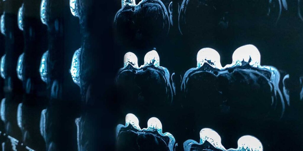
For women at high risk of breast cancer or who have dense breast tissue, a breast MRI is often recommended in addition to an annual mammogram. An MRI creates detailed pictures of the inner parts of the breast using radio waves and a strong magnetic field. Used together, a mammogram and a breast MRI can detect more cancers than just using one or the other. But the two imaging methods have important differences.
An MRI is not recommended as a screening tool on its own, because this type of imaging can miss some cancers that a mammogram would detect. An MRI is also more likely to return a false positive, or an abnormality that turns out to not be cancerous. False positives can lead to additional tests and biopsies that aren’t needed.
Doctors might recommend some women under the age of 40 begin getting a breast MRI before starting annual mammograms. Women in this group may include those with a strong family history of breast or ovarian cancer or certain genetic mutations that increase breast cancer risk.
Results, faster: accelerated breast MRI
The latest option for a breast MRI is the innovative accelerated breast MRI. This is a highly sensitive imaging method, showing more details than digital mammograms and taking less time than a standard MRI, usually 12 to 18 minutes. This type of imaging is a precise way to detect cancer in its earliest stages. In most cases, an accelerated breast MRI requires a contrast dye injection to help radiologists see the details of certain structures and interpret the results more accurately. This scan can also be done in an open MRI machine, providing additional comfort for patients.
What is a breast ultrasound?
Like a breast MRI, a breast ultrasound is not usually used as a screening method on its own. But when a mammogram detects something unusual or a lump can be felt in the breast, a breast ultrasound can give your doctor critical information.
Often, doctors order breast ultrasounds to determine whether a lump is filled with fluid, meaning it is likely a benign cyst, or if it is solid. Masses that are solid frequently need to be biopsied to determine whether they are cancerous or not. A surgeon can also use an ultrasound to help guide a biopsy needle into the correct area of the breast.
An ultrasound is usually performed with a handheld instrument called a transducer, which is moved over the skin of the area being imaged. The transducer works by sending sound waves and picking up echoes as they reflect off tissues deep under the skin. A special computer generates images based on these echoes.
Automated breast ultrasound (ABUS) for additional testing
A technique called automated breast ultrasound (ABUS) can be a good option for women with dense breast tissue. ABUS uses a much bigger transducer to take hundreds of pictures, covering nearly the entire breast. Doctors sometimes use ABUS for women who have an abnormal mammogram or have unusual symptoms.
American Health Imaging provides cost-effective, high-quality imaging services at convenient locations in Houston, Texas, and Georgia. If you are due for your annual screening mammogram or have a referral from your doctor for a diagnostic mammogram, breast MRI or breast ultrasound, request an appointment at an AHI location near you.
