
An MRI is the standard imaging test healthcare providers use to diagnose a torn ACL. If your doctor ordered a knee MRI, it’s because it gives them the detailed, accurate information they need, including the exact location and severity of the injury, whether it’s a partial or complete tear, and whether any other structures in the knee were affected at the same time.
This guide explains how MRI detects ACL tears, how accurate it is, what to expect during your appointment, and how American Health Imaging can help you get the answers you need fast and at a cost you can afford.
What Is the ACL and How Does It Get Torn?
The ACL, or anterior cruciate ligament, is one of four major ligaments stabilizing your knee. It connects the femur (thigh bone) to the tibia (shin bone) and controls how far the knee rotates and moves forward. Without a healthy ACL, your knee may feel unstable, especially when stopping suddenly, changing direction, or landing from a jump.
ACL tears are among the most common serious knee injuries, with an estimated 200,000 occurring in the United States every year. They happen most often during sports, like basketball, soccer, football and skiing, but can also result from non-athletic incidents that force the knee beyond its normal range of motion.
Symptoms of an ACL tear typically include a loud pop at the moment of injury, immediate swelling, pain, and a feeling that the knee might “give out.”
How Accurate Is an MRI for Diagnosing ACL Tears?
MRI scans are highly accurate for detecting ACL injuries. Published research shows MRI achieves approximately 95% sensitivity and 92–95% specificity for ACL tears overall. In practical terms, this means MRI correctly identifies the presence of a tear in the vast majority of cases.
Accuracy varies somewhat depending on technology and the nature of the tear:
- 3T MRI scanners achieve up to 95% accuracy even for partial ACL tears
Lower-field 1.5T scanners have reported accuracy of 25–53% for partial tears - Complete tears are generally easier to identify than partial tears on MRI
- Subspecialized musculoskeletal radiologists, those who read knee and joint MRIs primarily, produce more accurate reads than general radiologists
At American Health Imaging, all knee MRIs are read by board-certified, subspecialized radiologists with extensive experience in musculoskeletal imaging, giving you and your doctor the most accurate possible interpretation.
What Does a Torn ACL Look Like on MRI?
On a normal MRI, the ACL appears as a dark, taut band running diagonally through the knee joint. A torn ACL looks noticeably different. Common MRI findings that indicate an ACL tear include:
- A solid mass or irregular shape where continuous fibers should be visible
- Bright signal (white areas) within the ligament, indicating swelling or fluid
- A wavy or sagging appearance instead of a straight, taut band
- A visible gap or discontinuity in the ligament fibers
- Bone bruises on the lateral femoral condyle or tibial plateau (a secondary sign of ACL injury from impact)
Your radiologist will note these findings in the MRI report and describe the location and extent of the tear, giving your orthopedic provider the information they need to plan your care.
Partial vs. Complete ACL Tear: How MRI Tells the Difference
One of MRI’s key advantages is its ability to distinguish between a partial and a complete ACL tear — a distinction that significantly affects treatment decisions.
- Partial tear: Some fibers remain intact. The MRI may show thinning or irregular areas in the ligament, but some continuous fibers are still visible. The joint may remain relatively stable.
- Complete tear: The ligament has fully ruptured. MRI shows a distinct gap with no visible connection between the two ends. Joint instability is typically more pronounced.
ACL Tear Grades Explained (Grade I, II, III)
ACL injuries are often categorized into three grades based on severity:
- Grade I (Mild Sprain): The ACL has been stretched but not torn. Fibers are intact. The knee remains stable. MRI may show mild swelling around the ligament.
- Grade II (Partial Tear): Some ACL fibers have been torn. The ligament is partially intact but weakened. MRI shows disruption of some fibers with areas of increased signal intensity. The joint may feel loose or unstable.
- Grade III (Complete Rupture): The ACL is fully torn. MRI shows complete discontinuity of the ligament. This is the most common ACL injury requiring surgical consideration for active patients.
Your MRI report may reference these grades or describe findings using equivalent language. Your orthopedic provider will use the grade, along with your symptoms and activity level, to recommend the right treatment path.
What Else Can MRI Detect in an Injured Knee?
An ACL tear rarely happens in isolation. MRI is valuable not just for confirming the ACL injury but for revealing the full picture of what’s happening inside the knee. Co-injuries commonly seen on MRI alongside ACL tears include:
- Meniscus tears (most common, especially the lateral meniscus)
- Bone bruises or bone contusions on the femur and tibia from impact forces
- MCL (medial collateral ligament) damage
- PCL (posterior cruciate ligament) involvement in high-energy injuries
- Cartilage damage or early signs of wear
Identifying these associated injuries on MRI is critical for surgical planning. An ACL reconstruction performed without addressing a concurrent meniscus tear, for example, may not fully restore knee stability. Your orthopedic surgeon uses the complete MRI picture, not just the ACL finding, to make the most informed treatment decision.
MRI vs. Physical Exam: How ACL Tears Are Diagnosed
Your doctor likely performed a physical examination before ordering your MRI. Skilled clinicians can identify an ACL tear in up to 90% of cases through clinical tests like the Lachman test, anterior drawer test, and pivot shift test. So why order an MRI?
An MRI scan is ordered to:
- Confirm the tear and rule out other diagnoses
- Determine whether the tear is partial or complete
- Grade the severity and identify exact tear location for surgical planning
- Detect associated injuries (meniscus, cartilage, other ligaments) that physical exam alone cannot reliably identify
- Provide a pre-surgical baseline and legal documentation of the injury
In short, the physical exam points your doctor toward a diagnosis, while the MRI gives them the complete, accurate picture they need to treat you correctly.
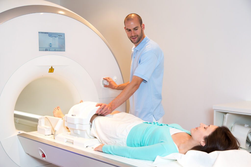
What to Expect During Your Knee MRI
How to Prepare
Preparing for a knee MRI is straightforward. Wear comfortable, loose-fitting clothing without metal (no zippers, no underwire). Remove jewelry, watches, and belts before your scan. Let your imaging team know in advance if you have any metal implants such as a pacemaker, surgical pins, or other hardware. In most cases, no fasting is required.
What Happens During the Scan
You’ll lie on a cushioned table that slides into the MRI machine. For a knee MRI, only your leg enters the machine and your head and upper body remain outside, which many patients find more comfortable than a full-body scan. The machine will make rhythmic thumping and buzzing sounds as it captures images. The scan itself is completely painless and non-invasive.
You’ll be asked to stay as still as possible during imaging. Your technologist will communicate with you throughout via intercom. If you feel uncomfortable at any point, let them know; they can pause and reposition you if needed.
How Long Does a Knee MRI Take?
Most knee MRI appointments take less than 30 minutes total. At most American Health Imaging centers, faster scanning technology reduces your actual time inside the scanner to an average of 15 minutes or less — up to 50% faster than standard equipment.
How Your Doctor Uses MRI Results to Plan Your Treatment
Your MRI findings directly influence whether your provider recommends surgery, physical therapy, or a combination of both:
- Partial tears (Grade I–II): Many partial ACL tears, especially in less active patients or older adults, can be managed successfully with physical therapy, bracing, and activity modification. MRI findings showing intact fibers and a stable joint support a non-surgical approach.
- Complete tears (Grade III): Athletes, younger patients, and anyone wanting to return to high-demand physical activity typically benefit from surgical ACL reconstruction. MRI confirms the complete rupture and guides the surgical plan.
Schedule Your Knee MRI at American Health Imaging
Choosing clarity about your knee health starts with choosing the right imaging provider. American Health Imaging combines advanced MRI technology with board-certified, subspecialized musculoskeletal radiologists to deliver the precise, expert reads your doctor needs to treat you correctly.
We offer same-day and next-day appointments, extended hours, and convenient locations across Georgia, Alabama, Texas, South Carolina, and Florida, with results delivered to you and your doctor within 48 hours.
Talk to your doctor about scheduling your knee MRI with American Health Imaging today.
Frequently Asked Questions About MRI Scans for ACL Tears
After your scan, a board-certified, subspecialized radiologist reviews your images and sends a detailed report to your doctor. At American Health Imaging, we also text you a link to an easy-to-read imaging report within 48 hours, with clear explanations and helpful diagrams so you can understand your results before your follow-up appointment.
Your orthopedic provider will then review the results with you, explain the findings, and discuss your treatment options.
Recovery timelines vary based on your MRI findings and treatment choice:
- Non-surgical management: 3–6 months of physical therapy with gradual return to activity
- ACL reconstruction surgery: Typically 6–9 months before return to sport, and sometimes longer for high-level athletic demands
Your MRI results, including associated injuries like meniscus tears or cartilage damage, directly affect your recovery timeline and rehabilitation plan. This is why a thorough, expert MRI read is so important from the very start.
Cost is one of the most common questions patients have about knee MRI. The price of a knee MRI varies widely depending on where you go. Hospital-based imaging centers typically charge significantly more than independent imaging providers like American Health Imaging.
American Health Imaging offers knee MRI at up to 60% less than hospital-based pricing. We are in-network with 99% of insurance plans, and our team can help you understand your benefits and out-of-pocket costs before your appointment, so there are no surprises.
If you have a high-deductible plan, booking directly with an independent imaging center like AHI is often significantly less expensive than using a hospital-based option even after insurance.
An MRI creates detailed images of the soft tissues inside your knee, allowing your radiologist to see the ACL's fibers directly. A torn ACL shows fraying, gaps, swelling, or complete separation of the ligament — findings that a physical exam and X-ray cannot reliably detect.
MRI is approximately 95% sensitive and 92–95% specific for ACL tears. It is the most accurate non-invasive diagnostic tool available and is considered the standard of care before any surgical decision for an ACL injury.
ACL injuries are graded Grade I (mild stretch, ligament intact), Grade II (partial tear, some fibers damaged), and Grade III (complete rupture). MRI helps determine the grade, which directly influences whether treatment is surgical or non-surgical.
Yes. A partial tear shows intact but irregular or thinned fibers, while a complete tear shows a visible gap with no connection between the torn ends. This distinction is critical for treatment planning.
No. For a knee MRI, only your leg enters the scanner. Your head and upper body remain outside the machine, which most patients find much more comfortable than scans that require full-body entry.