American Health Imaging of Sandy Springs

Fax:

404-963-0632

Hours

Mon-Fri: 7:00am - 10:00pm
Sat: 8:00am - 2:00pm
Sun: Closed

Services

  • Accelerated Breast MRI
  • Arthrogram
  • Cardiac Scoring
  • CT
  • Diffusion Tensor Imaging
  • Low-Dose CT Lung Cancer Screening
  • MRI
  • Myelogram
  • Ultrasound
American Health Imaging Outpatient Medical Diagnostic Imaging in the Sandy Springs area in Atlanta, Georgia

Welcome to American Health Imaging of Sandy Springs in Georgia

American Health Imaging (AHI) of Sandy Springs is proud to be part of the AHI network with 22 centers throughout the Atlanta metroplex, bringing state-of-the-art medical imaging services to Sandy Springs, Georgia. We are conveniently located on Lake Hearn Drive, near the intersection of Peachtree Dunwoody Road and Johnson Ferry Road. AHI Sandy Springs offers MRI, CT, and Ultrasound exams, as well as specialty scans including calcium cardiac scoring, low dose lung cancer screening, fluoroscopy, and arthrogram.

AHI of Sandy Springs offers same-day and next-day appointments and early morning, evening, and weekend hours so you can get your imaging service when it fits with your busy schedule. AHI costs up to 60% less than hospital based imaging and is in-network with 99% of major health insurance plans, providing a cost-effective option for your diagnostic imaging needs. Schedule your appointment with AHI Sandy Springs today.

Medical Imaging Services Offered at Atlanta, Georgia

Services at This Location:

Tap services to view more information

A shorter, radiation-free breast MRI that provides more detail than a mammogram alone, which can be especially helpful for women with dense breast tissue.
A contrast-enhanced MRI or X-ray exam of a joint that shows tendons, ligaments, cartilage and other structures in detail, helping pinpoint the cause of joint pain.
A quick, non-invasive CT scan that measures calcium buildup in your heart’s arteries to help gauge your risk of coronary artery disease before symptoms appear.
CT uses specialized X-rays to create cross-sectional images of bones, organs and blood vessels, helping your doctor quickly diagnose injuries, infections and other conditions.
An advanced MRI technique that maps the brain’s white matter pathways, helping doctors assess the effects of traumatic brain injuries.
A low-dose CT scan of the chest that looks for early signs of lung cancer in people at higher risk, when it’s often easier to treat.
Combines a contrast injection with imaging to show the spinal cord, nerve roots and discs in detail, helping diagnose back pain, nerve compression and other spine problems.
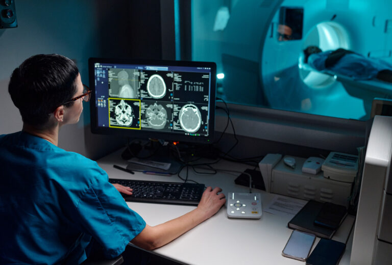
MRI uses powerful magnets and radio waves to create detailed images of the brain, spine, joints, organs and soft tissues without radiation. It helps your doctor diagnose injuries and health conditions and see how well a treatment is working.
Ultrasound uses sound waves to create real-time images of organs, blood vessels and soft tissues, without radiation.

Advanced Imaging Technology at Our Atlanta Center

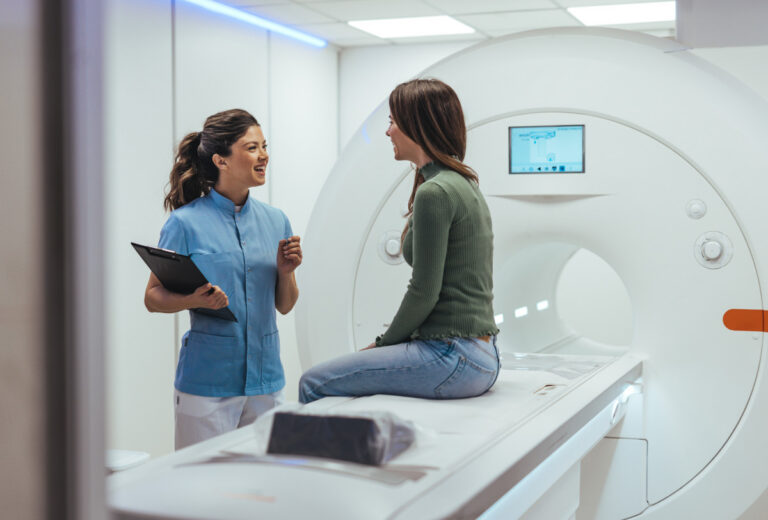
A 1.5T Wide Bore MRI is a type of medical imaging machine that uses a strong magnetic field (1.5 Tesla) to take detailed pictures of the inside of your body. The “wide bore” part means the opening of the machine is larger at 27 ½” wide, making it more comfortable for people who feel anxious or are larger in size. Many of our 1.5T Wide Bore MRIs feature faster scanning technology to reduce most exam times to as little as 8 minutes.

A 3T MRI is a medical imaging machine that uses an even stronger magnetic field (3 Tesla) to take very detailed pictures of the inside of your body. Because of its high strength, it can capture clearer images and is often used for more complex scans. Often used for prostates and different types of brain imaging. The “wide bore” part means the opening of the machine is larger at 27 ½” wide, making it more comfortable for people who feel anxious or are larger in size. Available at select AHI imaging centers.

A 64-slice CT scanner is a medical imaging machine that takes very detailed X-ray pictures of your body by capturing 64 slices of images at once. This allows for faster scans and even more detailed images, which is useful for diagnosing complex conditions. Our 64-slice CT features innovative technology that automates dose according to your size, weight, and anatomy, providing high-quality images with minimal radiation.

Faster scanning MRI technology reduces the time patients spend on the table for scans by up to 50% to an average scan time of less than 15 minutes providing a more comfortable imaging experience for anxious patients or anyone in pain. AI technology delivers high quality images with reduced motion artifacts and noise distortions for the diagnostic insights providers need to determine next steps in patient care.

Fluoroscopy uses injected contrast dye and an X-Ray machine to take a continuous series of X-rays instead of individual snapshots. It is most commonly used to evaluate parts of your body that are moving in order to create a short video of your body system in motion. It is particularly useful for observing the digestive, urinary, respiratory, and reproductive systems and their functioning.

Ultrasound, also known as sonography, has a small handheld device called a transducer used to emit high-frequency sound waves into the body making it particularly useful for examining developing fetuses during pregnancy and for imaging soft tissues and organs.

Patient Reviews and Testimonials

With a 97% satisfaction score, we’re proud to deliver compassionate care, clear communication, and imaging you can count on.


Have you visited a American Health Imaging Center lately?

Medical Imaging Articles and Resources