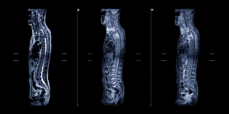


Accelerated Breast MRI (also called abbreviated breast MRI or AB-MRI) is a shortened, streamlined version of a traditional breast MRI that takes just 10-15 minutes while providing the same exceptional cancer-detection capabilities. This fast, radiation-free imaging test uses powerful magnets and radio waves to create detailed images of breast tissue, detecting cancers that mammograms often miss.
Key differences from traditional breast MRI:
• Scan time: 10-15 minutes (vs. 30-45 minutes for traditional breast MRI)
• Protocol: Focuses on essential sequences for cancer detection
• Cost: More affordable due to shorter scan time
• Detection: Same excellent cancer-detection rates
Accelerated Breast MRI is ideal for:
• Women with dense breasts (where mammography has limited sensitivity)
• Women at increased risk of breast cancer
• Supplemental screening alongside annual mammography
• Women with personal or family history of breast cancer
Your doctor ordered an Accelerated Breast MRI because you need the most sensitive breast cancer screening available, particularly if you have dense breasts or elevated breast cancer risk.
Common reasons your doctor ordered this test:
• Dense breasts: Dense tissue appears white on mammograms, and so do cancers, making tumors difficult to see
• High cancer risk: Family history, genetic mutations (BRCA1/BRCA2), or personal history of breast cancer
• Supplemental screening: To catch cancers that mammography might miss
• Follow-up imaging: Additional evaluation after an abnormal mammogram or ultrasound
• Implant evaluation: To check breast implants for rupture or leakage
The benefit: Studies show that breast MRI reduces interval cancers (cancers found between screenings) by more than 50% in women with extremely dense breasts.
Yes, you need a referral (also called a doctor’s order or prescription) to get an Accelerated Breast MRI scan.
What your referral does:
• Is based on your breast density, risk factors, and medical history
• Is necessary for insurance coverage
• Ensures the exam is appropriate for your specific situation
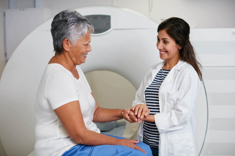
Once your doctor sends the order to American Health Imaging, we’ll contact you to schedule your appointment and verify your insurance coverage.
Scheduling a same-day Accelerated Breast MRI scan at American Health Imaging is easy and convenient.
Flexible scheduling options:
• Call us or request an appointment online
• Same-day and next-day appointments available
• Convenient hours to fit your schedule
Once your doctor sends us your referral, we can often schedule your scan quickly at one of our Georgia locations offering the scan.
American Health Imaging offers Accelerated Breast MRI at three convenient locations in Georgia.
Our Accelerated Breast MRI locations:
• Decatur, Georgia
• Lawrenceville, Georgia
• Sandy Springs, Georgia
All locations offer same-day and next-day appointments with convenient hours, ample parking, and compassionate, experienced staff. To find the location nearest you or schedule your appointment, call one of our imaging centers offering ABMRI or use our online scheduling system.
View All Imaging Centers
Accelerated Breast MRI is the most sensitive breast imaging test available for detecting breast cancer, particularly in women with dense breasts.
Conditions diagnosed with Accelerated Breast MRI:
• Breast cancer: Detects invasive cancers and some types of DCIS (ductal carcinoma in situ)
• Early-stage tumors: Finds cancers as small as 2-3 millimeters
• Breast implant problems: Evaluates silicone implants for rupture or leakage
• Breast abnormalities: Detects suspicious areas that need further evaluation
Detection rates:
• Sensitivity of 90-100% for detecting breast cancer
• Detects 15-16 additional cancers per 1,000 screenings compared to mammography alone
• Finds cancers at earlier, more treatable stages
• More than 40% of MRI-detected cancers are smaller than 1 cm
Accelerated Breast MRI is recommended for women who need supplemental breast cancer screening beyond mammography.
Women with dense breasts:
• Heterogeneously dense or extremely dense breast tissue
• Dense tissue makes mammograms less effective at detecting cancer
• About 40-50% of women have dense breasts
Women at high risk for breast cancer:
• BRCA1 or BRCA2 gene mutations
• Strong family history of breast or ovarian cancer
• Lifetime breast cancer risk of 20% or greater
• Personal history of breast cancer, especially if diagnosed before age 50
• Previous chest radiation between ages 10-30
• Genetic syndromes (Li-Fraumeni, Cowden, PTEN)
Women with specific breast conditions:
• Atypical hyperplasia or lobular carcinoma in situ (LCIS)
• Silicone breast implants (recommended every 2-3 years to check for rupture)
Talk to your doctor about whether Accelerated Breast MRI is right for you based on your individual risk factors and breast density.
Preparing for an Accelerated Breast MRI requires some simple steps to ensure the best images.
Before your appointment:
• Schedule your exam for the week after your period, when breasts are least tender (if you’re still menstruating)
• Inform us if you’re pregnant, breastfeeding, or might be pregnant
• Tell us about any kidney problems or previous reactions to contrast dye
• Arrive 15 minutes early to complete paperwork
Day of your appointment:
• Don’t wear deodorant, powder, lotion, or perfume on your chest or underarms (can interfere with imaging)
• Wear comfortable, two-piece clothing (you’ll change into a gown)
• Remove all jewelry, watches, and metal objects
Tell the technologist if you have:
• Metal implants, pacemaker, or IUD
• Breast implants (silicone or saline)
• Claustrophobia or anxiety
• Tattoos (some inks contain metal)
• Previous breast surgeries or biopsies
• Known allergies to contrast dye
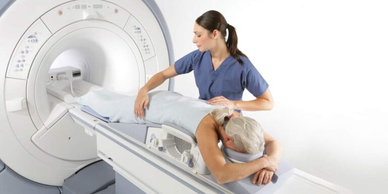
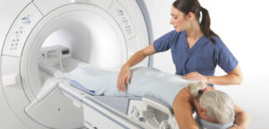
During your Accelerated Breast MRI scan, you’ll lie face-down with your breasts positioned comfortably in cushioned openings. The entire scan takes just 10-15 minutes.
What happens during your scan:
• Check-in: Arrive 15 minutes early; verify information and complete forms
• Screening: Answer safety questions about metal implants, kidney function, and allergies
• IV placement: A small IV placed in your arm for contrast injection
• Positioning: You’ll lie face-down with breasts in cushioned openings, arms extended above head
• Comfort measures: Pillows, blankets, and headphones with music provided
• The scan: Table slides into MRI machine (open at both ends)
• Contrast injection: Contrast dye injected partway through (you may feel warmth or cool sensation)
• Duration: 10-15 minutes total scan time
• Communication: You can talk to the technologist through an intercom anytime
Key benefits of Accelerated Breast MRI:
• Significantly shorter than traditional 30-45 minute breast MRI
• Open at both ends—less claustrophobic
• Same excellent cancer detection capability
After your Accelerated Breast MRI, you can resume all normal activities immediately.
Immediately after your scan:
• Resume driving, working, eating, and exercising
• Drink plenty of water for the rest of the day to help flush the contrast
• You can eat and take medications normally
• The IV site may be slightly tender or bruised (normal and temporary)
Getting your results:
• Fellowship-trained breast imaging radiologists review your images
• Your doctor receives a complete written report
• You’ll receive a text message with a link to a patient-friendly version of your report with explanations and diagrams
• Your doctor will contact you to discuss findings and determine if any follow-up is needed
Good news: Most breast MRI results are normal. If an abnormality is detected, your doctor may recommend additional imaging or a biopsy.
After your exam results are finalized, your doctor will receive them and discuss your findings with you. You will receive a patient-friendly report via text 48 hours after the report is finalized.
Reports are sent via our Scanslated patient report system and provides easy-to-understand explanations of your scan and diagrams. View your report anytime on your computer or mobile device once sent.
What’s included in your patient report:
• Easy-to-understand explanations of medical terminology
• BI-RADS category (standardized reporting system)
• Anatomical diagrams
• Information to help you prepare for your follow-up conversation with your doctor
Your breast MRI images are reviewed by fellowship-trained breast imaging radiologists who specialize in detecting breast cancer.
Accelerated Breast MRI is the most sensitive breast imaging test available for detecting breast cancer.
Accuracy and detection rates:
• Sensitivity: 90-100% for detecting breast cancer
• Far superior to mammography: Especially in dense breasts (mammography: 25-59% in dense breasts)
• Detects more cancers: 15-16 additional cancers per 1,000 screenings compared to mammography alone
• Finds smaller cancers: More than 40% are under 1 cm (highly curable)
• Earlier detection: MRI-detected cancers are less likely to have spread to lymph nodes
Research shows:
• Reduces interval cancer rates by more than 50%
• Detects cancers at earlier, more treatable stages
• Maintains the same cancer detection rates as traditional full-protocol breast MRI
Important to know: Breast MRI has a higher false-positive rate than mammography (may find suspicious areas that turn out to be benign). This is why breast MRI is recommended as a supplement to—not a replacement for—annual mammography.
The actual scan: Just 10-15 minutes from start to finish
Your total appointment time: 30-40 minutes (including check-in, changing clothes, IV placement, and post-scan instructions)
Why it’s faster:
• Streamlined protocol focuses on essential cancer-detection sequences
• Significantly shorter than traditional breast MRI (30-45 minutes)
• Makes breast MRI more comfortable and convenient
The cost of Accelerated Breast MRI varies depending on your insurance coverage and whether you’re paying out-of-pocket.
Typical Accelerated Breast MRI costs:
• With insurance: Depends on your deductible, coinsurance, and coverage for your specific indication
• Self-pay rates: Up to 60% less than hospital pricing
• More affordable: Than traditional breast MRI due to shorter scan time
We’re in-network with 99% of insurance plans, which means lower out-of-pocket costs for you.
What we do for you:
• Verify your insurance coverage
• Obtain necessary pre-authorization
• Provide a cost estimate before your appointment
• Offer transparent self-pay rates if insurance doesn’t cover
American Health Imaging can save patients hundreds to thousands of dollars compared to hospital-based breast MRI while providing the same high-quality imaging and expert interpretation.
Insurance coverage for Accelerated Breast MRI varies by plan and depends on your specific risk factors and indication for the exam.
Generally covered when:
• You have a BRCA1 or BRCA2 gene mutation
• You have a lifetime breast cancer risk of 20% or greater
• You have a personal history of breast cancer
• You have certain genetic syndromes (Li-Fraumeni, Cowden)
• You have a first-degree relative with BRCA mutations
• Your doctor orders it for evaluation of breast implants
May require pre-authorization for:
• Dense breasts as the sole indication
• Intermediate-risk patients (15-20% lifetime risk)
• Supplemental screening purposes
American Health Imaging is in-network with 99% of insurance plans. We’ll verify your coverage, obtain necessary pre-authorization, and provide a cost estimate before your appointment. If insurance doesn’t cover your scan, we offer transparent self-pay rates.
Dense breast tissue means you have more glandular and fibrous tissue and less fatty tissue in your breasts.
Breast density categories:
• A – Almost entirely fatty: Breasts are mostly fat
• B – Scattered fibroglandular density: Some dense tissue scattered throughout
• C – Heterogeneously dense: Breasts have many areas of dense tissue (about 40% of women)
• D – Extremely dense: Breasts are mostly dense tissue (about 10% of women)
Why breast density matters:
1. Increases breast cancer risk: Women with dense breasts are 4-6 times more likely to develop breast cancer
2. Masks cancers on mammograms: Both dense tissue and cancers appear white on mammograms, making tumors difficult to see
3. Increases interval cancer risk: More likely to develop cancers between screenings
What to do: After your mammogram, you’ll receive a letter indicating your breast density category. If you have heterogeneously dense or extremely dense breasts, ask your doctor about supplemental screening with Accelerated Breast MRI.
Accelerated Breast MRI uses a shortened scanning protocol while maintaining the same excellent cancer-detection capabilities.
Key Differences: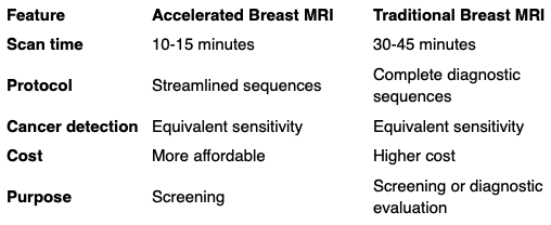
Studies show: Accelerated Breast MRI maintains the same excellent cancer-detection rates as traditional breast MRI while being faster, more comfortable, and more cost-effective for screening.
Important: If an abnormality is detected on your Accelerated Breast MRI, you may need a full diagnostic breast MRI for complete characterization.
Accelerated Breast MRI and mammography serve complementary roles—they work best together, not as replacements for one another.
Breast MRI advantages:
• Much higher sensitivity for detecting cancer (90-100% vs. 25-59% for mammography in dense breasts)
• Detects cancers at earlier stages
• Not limited by breast density—can “see through” dense tissue
• Finds cancers that mammograms miss
• Reduces interval cancers by more than 50%
• No radiation exposure
Mammography advantages:
• Excellent at detecting microcalcifications (early signs of DCIS)
• Widely available and less expensive
• Faster and more convenient
• Lower false-positive rate
• Established mortality benefit in randomized trials
The ideal approach: For women with dense breasts or high risk, annual mammography PLUS annual supplemental breast MRI provides the most comprehensive screening available.
Contrast dye (gadolinium) is essential for breast MRI because it highlights areas of abnormal blood vessel growth, a hallmark of breast cancer.
How contrast works:
• Breast cancers create new blood vessels to fuel their growth
• These abnormal vessels are “leaky”
• Contrast agent accumulates in these areas
• Causes breast cancers to “light up” on MRI images
• Allows detection of cancers as small as 2-3 millimeters
Important: Without contrast, breast MRI cannot reliably detect breast cancer.
Yes! In fact, breast MRI is the gold standard for evaluating silicone breast implants for rupture or leakage.
MRI for breast implants:
• The FDA recommends women with silicone implants undergo breast MRI every 2-3 years
• Detects silent ruptures (ruptures without symptoms)
• Evaluates implant positioning and integrity
• Identifies leaking silicone
• Assesses surrounding breast tissue for cancer
Important to know:
• Breast implants do not interfere with the MRI scan
• The MRI magnetic field does not damage or affect your implants
• Tell the technologist about your implants so they can use appropriate imaging sequences
If your Accelerated Breast MRI detects a suspicious area, don’t panic—most findings turn out to be benign after additional evaluation.
BI-RADS categories your radiologist will assign:
• BI-RADS 0: Additional imaging needed (callback for more views)
• BI-RADS 1: Negative (no abnormalities)
• BI-RADS 2: Benign findings (no follow-up needed)
• BI-RADS 3: Probably benign (short-term follow-up MRI in 6 months recommended)
• BI-RADS 4: Suspicious (biopsy recommended)
• BI-RADS 5: Highly suggestive of cancer (biopsy required)
Remember: Early detection saves lives. Finding something on MRI often means catching cancer at its earliest, most treatable stage.
The frequency of breast MRI screening depends on your risk level and breast density.
Annual breast MRI recommended for:
• Women with BRCA1 or BRCA2 mutations
• Women with 20% or greater lifetime breast cancer risk
• Women with a personal history of breast cancer (especially if dense breasts)
• Women with certain genetic syndromes
• Women who had chest radiation between ages 10 and 30
Consider annual breast MRI for:
• Women with extremely dense breasts
• Women with intermediate risk (15-20% lifetime risk)
• Women with strong family history of breast cancer
• Women with atypical hyperplasia or LCIS
Timing: Most women alternate their MRI and mammogram every 6 months (MRI in spring, mammogram in fall), so they’re being screened twice yearly with different modalities. Some women have both tests done on the same day.
Talk to your doctor about the screening schedule that’s right for you.
Yes, Accelerated Breast MRI is very safe. MRI uses powerful magnets and radio waves—no radiation—making it safer than mammography or CT scans for repeated imaging.
Safety benefits:
No radiation exposure: Unlike mammograms and CT scans
• Contrast safety: Gadolinium contrast is very safe; serious allergic reactions occur in less than 0.01% of patients
• Safe for repeated screening: No cumulative radiation risk
Who should not have a breast MRI:
• Patients with certain metal implants (some pacemakers, cochlear implants)
• Patients with metal fragments in their eyes
• Patients with severe kidney disease (contrast may not be safe)
• First trimester of pregnancy (although no harm has been proven)
Always inform your technologist about any metal in your body, kidney problems, or allergies before your MRI.
Yes—this is one of the most important benefits of Accelerated Breast MRI.
Evidence of early detection:
• More than 40% of MRI-detected cancers are smaller than 1 cm (highly curable)
• MRI-detected cancers are less likely to have spread to lymph nodes (21% vs. 55% for mammography-detected cancers)
• Interval cancer rates reduced by more than 50% with MRI screening
• Earlier detection translates to better survival rates and less aggressive treatment
For high-risk women: Studies show that adding annual MRI to mammography screening significantly improves survival compared to mammography alone.
The benefit: Early detection means smaller tumors, less invasive surgery, better cosmetic outcomes, and higher cure rates.