Diffusion Tensor Imaging

What is Diffusion Tensor Imaging?

Diffusion Tensor Imaging (DTI) is an advanced type of MRI brain scan that maps the brain’s white matter pathways with remarkable precision. Unlike standard MRI scans that show brain structure, DTI tracks water movement along nerve fibers to reveal the integrity and organization of white matter tracts, the neural highways connecting different brain regions. DTI can detect damage from traumatic brain injury, stroke, multiple sclerosis, or degenerative disease, even when a standard MRI appears normal.

DTI is particularly valuable for:

  • Brain tumor surgical planning and protecting critical pathways
  • Traumatic brain injury and concussion assessment
  • Stroke recovery prediction and rehabilitation planning
  • Multiple sclerosis diagnosis and monitoring
  • Alzheimer’s and Parkinson’s disease evaluation

Diffusion Tensor Imaging Services at American Health Imaging

American Health Imaging is in-network with 99% of insurance plans and offers DTI brain imaging up to 60% less than hospital-based imaging.

Diffusion Tensor Imaging Centers Near You

Find convenient DTI imaging at select American Health Imaging centers in Alabama, Georgia, Texas, Florida, and South Carolina. We offer same-day and next-day appointments with extended hours and weekend availability to match your schedule and needs.

Find Your Nearest Diffusion Tensor Imaging Imaging Center

What is Diffusion Tensor Imaging used for?

Diffusion Tensor Imaging (DTI) provides valuable diagnostic information that cannot be obtained from standard MRI scans. DTI is particularly valuable when doctors need to assess white matter integrity, plan brain surgery around critical pathways, or detect subtle brain injuries invisible on conventional imaging. Doctors use DTI to diagnose traumatic brain injury, guide tumor surgery, evaluate stroke damage, monitor multiple sclerosis, and assess neurodegenerative diseases—all without radiation exposure.

Common DTI Scans and Uses:

  • Brain Tumor Surgical Planning
  • Traumatic Brain Injury (TBI) and Concussion
  • Stroke Evaluation and Recovery Prediction
  • Multiple Sclerosis Diagnosis and Monitoring
  • Alzheimer’s and Neurodegenerative Diseases
  • Epilepsy Presurgical Evaluation
  • Pediatric Developmental Disorders

Types of Diffusion Tensor Imaging Scans We Offer

A 1.2T High Field Open MRI is a type of Open MRI scanner that has a strong magnet (1.2 Tesla) and an open design, making it more comfortable for people who feel claustrophobic or have a larger body. The Open MRI has an open front and sides designed to provide increased comfort. The “high field” means it has a stronger magnetic field than some other MRI machines, which helps in getting more accurate images.

A 1.5T High Field MRI is a medical imaging machine that uses a strong magnetic field (1.5 Tesla) to create clear and detailed pictures of the inside of your body. The “high field” means it has a stronger magnetic field than some other MRI machines, which helps in getting more accurate images.

A 1.5T Wide Bore MRI is a type of medical imaging machine that uses a strong magnetic field (1.5 Tesla) to take detailed pictures of the inside of your body. The “wide bore” part means the opening of the machine is larger at 27 ½” wide, making it more comfortable for people who feel anxious or are larger in size. Many of our 1.5T Wide Bore MRIs feature faster scanning technology to reduce most exams to 15 minutes.

A 16-slice CT scanner is a medical imaging machine that takes detailed X-ray pictures of your body in thin slices, which are then combined to create a complete image. The “16-slice” part means it can capture 16 slices of images at once, allowing for faster and more detailed scans.

3D mammography, or tomosynthesis, is an advanced x-ray technology that takes multiple images of breast tissue to create a 3-dimensional picture of the breast. It differs from traditional mammography in that traditional mammography yields only a single image. A 3D image of the breast allows for better assessment of masses and reduces the likelihood of a false positive or an unnecessary biopsy.

A 3T MRI is a medical imaging machine that uses an even stronger magnetic field (3 Tesla) to take very detailed pictures of the inside of your body. Because of its high strength, it can capture clearer images and is often used for more complex scans. Often used for prostates and different types of brain imaging. The “wide bore” part means the opening of the machine is larger at 27 ½” wide, making it more comfortable for people who feel anxious or are larger in size.

A 64-slice CT scanner is a medical imaging machine that takes very detailed X-ray pictures of your body by capturing 64 slices of images at once. This allows for faster scans and even more detailed images, which is useful for diagnosing complex conditions. Our 64-slice CT features innovative technology that automates dose according to your size, weight, and anatomy, providing high-quality images with minimal radiation.

ABUS Breast Cancer Screening uses sound waves to create 3D pictures of the breast tissue. ABUS breast cancer screening technology is specifically developed and FDA-approved for women with dense breast tissue.

An arthrogram visualizes the inside of a joint, such as the shoulder, knee, hip, or wrist. A contrast dye is injected into the joint to make the joint structures, including ligaments, tendons, cartilage, and the joint capsule, more visible on X-ray or MRI images.

Coronary Calcium CT – also called Cardiac Scoring – measures calcified (hard) plaque inside the arteries that can grow and restrict blood flow to the muscles of the heart identifying potential Coronary Artery Disease (CAD) before you have symptoms.

A DEXA scan is a medical imaging technique used to measure bone mineral density. It is commonly performed to diagnose osteoporosis or assess the risk of developing fractures as we age.

Diffusion Tensor Imaging (DTI) is a special type of MRI technique that helps doctors see the pathways of nerve fibers in the brain. By looking at these images, doctors can better understand how the brain’s wiring works and diagnose conditions like brain injuries, tumors, or diseases that affect these connections.

Faster scanning MRI technology reduces the time patients spend on the table for scans by up to 50% to an average scan time of less than 15 minutes providing a more comfortable imaging experience for anxious patients or anyone in pain. AI technology delivers high quality images with reduced motion artifacts and noise distortions for the diagnostic insights providers need to determine next steps in patient care.

Fluoroscopy uses injected contrast dye and an X-Ray machine to take a continuous series of X-rays instead of individual snapshots. It is most commonly used to evaluate parts of your body that are moving in order to create a short video of your body system in motion. It is particularly useful for observing the digestive, urinary, respiratory, and reproductive systems and their functioning.

A screening mammography is a specialized X-ray of the breasts that takes images from different angles. These images can reveal abnormalities in breast tissues. This exam is annually recommended for all women over the age of 40.

Our MRI machines use a powerful magnetic field, radio waves, and a computer to generate detailed images of organs, tissues, bones, and other structures inside the body. With a larger bore or tunnel-like opening compared to traditional MRI machines, our wide-bore MRI provides high quality images without compromising patient comfort.

Our MRI machines use a powerful magnetic field, radio waves, and a computer to generate detailed images of organs, tissues, bones, and other structures inside the body. With a larger bore or tunnel-like opening compared to traditional MRI machines, our wide-bore MRI provides high quality images without compromising patient comfort.

Myelogram imaging is used to evaluate the spinal cord, nerve roots, and surrounding structures in the spine with an injectable contrast dye to enhance the visibility of the spinal cord and nerves on X-ray images.

Specialty post processing technology used for brain MRI to assess brain volume and atrophy for neurological diseases including dementia, Alzheimers, seizures, MS lesions and Traumatic Brain Injuries.

The Open Upright MRI, also known as a stand-up MRI, is the only MRI scanner able to scan you in multiple positions, including sitting, standing, bending (for flexion and extension) or lying down. This unique MRI provides natural weight-bearing imaging and is helpful for your doctor to diagnose the area where you experience pain. The Open Upright MRI is open in front of you, behind you, and above you. This open design may be more comfortable for people who feel anxious or are larger in size.

A PET/CT (Positron emission tomography) is a non-invasive exam that uses small amounts of radioactive materials called radiotracers to reveal how internal organs are functioning. The scan takes about 60 minutes and images are captured via CT as your body processes the radioactive material. This offers great insight into the root of the disease process or the source of symptoms.

What to Expect During Your Diffusion Tensor Imaging Appointment

Your Diffusion Tensor Imaging (DTI) appointment begins with a standard brain MRI that includes one additional specialized sequence lasting about 4–5 minutes. You’ll lie comfortably on the scanning table as it moves into the MRI machine, and it’s especially important to stay completely still during the DTI portion so the system can accurately measure your brain’s white matter pathways. Our imaging technologists will guide you through the process and check in on your comfort throughout the exam.

Most DTI scans take 30–60 minutes in total. You’ll hear rhythmic thumping and clicking sounds during imaging, completely normal, and we’ll provide headphones with music or earplugs to help you relax.

Why Choose American Health Imaging for Your Diffusion Tensor Imaging

American Health Imaging (AHI) is in-network with 99% of insurance plans for DTI imaging. It offers compassionate care combined with state-of-the-art technology for accuracy and results you can trust.

  • Significant cost savings up to 60% compared to hospital DTI pricing
  • Advanced 1.5T and 3T MRI scanners with multi-directional gradient sequences for superior white matter mapping
  • board-certified, subspecialty neuroradiologists read DTI exams for accurate interpretations you and your doctor can rely on
  • Scanslated® patient-friendly reports help you understand your results
  • With a 97% patient satisfaction score, patients love our compassionate staff and comfortable imaging experience

Your health matters, and so does your time. That’s why we offer same-day and next-day appointments, extended hours, and weekend availability at convenient locations across the Southeast.

Schedule Your DTI Scan Today

Getting started is easy:

  1. Get a referral from your doctor (required for insurance coverage)
  2. Confirm DTI availability at your preferred American Health Imaging location—call ahead, as not all centers offer this specialized imaging
  3. Schedule your appointment online or by phone—same-day slots often available
  4. Verify insurance coverage—we’ll handle pre-approval and certification

Common Questions About Diffusion Tensor Imaging Scans

What is a DTI scan?

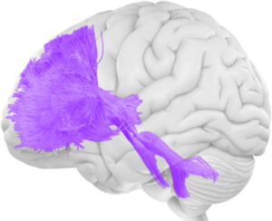
DTI (Diffusion Tensor Imaging) is an advanced type of MRI brain scan that maps the brain’s white matter pathways with remarkable precision. Unlike standard MRI scans that show brain structure, DTI tracks water movement along nerve fibers to reveal the integrity and organization of white matter tracts—the neural highways connecting different brain regions.

How DTI works:
• Uses magnetic fields and radio waves (no radiation)
• Measures microscopic water movement in brain tissue
• Tracks water diffusion along nerve fiber bundles
• Creates 3D maps of white matter pathways (fiber tractography)
• Produces color-coded images showing brain connectivity

Key benefits:
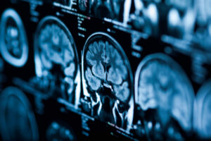
• Detects brain damage invisible on standard MRI
• Maps critical pathways before brain surgery
• Predicts stroke recovery with up to 80% accuracy
• Monitors disease progression over time
• Non-invasive and painless

DTI is particularly valuable for:
• Brain tumor surgical planning and protecting critical pathways
• Traumatic brain injury and concussion assessment
• Stroke recovery prediction and rehabilitation planning
• Multiple sclerosis diagnosis and monitoring
• Alzheimer’s and Parkinson’s disease evaluation
• Epilepsy presurgical evaluation
• Pediatric developmental disorders (autism, ADHD, cerebral palsy)

What makes DTI different from regular MRI:
Think of a standard MRI as a photograph of a highway system, while DTI shows which lanes are open, which are blocked, and how traffic flows.

Why did my doctor order a DTI scan?

Your doctor ordered a DTI scan to evaluate white matter integrity, brain connectivity, or neural pathway damage that cannot be seen on standard MRI scans. DTI provides critical information for diagnosis, treatment planning, and monitoring neurological conditions.

Common reasons your doctor ordered this test:
• Brain injury assessment (concussion or traumatic brain injury)
• Brain tumor evaluation and surgical planning
• Stroke evaluation and recovery prediction
• Neurological disease monitoring
• Unexplained neurological symptoms
• Pediatric neurological conditions
• Presurgical evaluation

The benefit:
DTI reveals white matter damage and neural pathway disruption that would remain invisible on conventional MRI, leading to more accurate diagnoses and better treatment planning.

Do I need a referral for a DTI scan?

Yes, you need a referral (also called a doctor’s order or prescription) to get a DTI scan at American Health Imaging.

What your referral does:
• Specifies that DTI imaging is medically necessary
• Provides clinical information about your symptoms and condition
• Is required for insurance coverage
• Ensures the appropriate imaging protocol is performed
• Allows the radiologist to provide targeted interpretation

What information your doctor provides:
• Specific clinical indication (TBI, stroke, tumor, MS, etc.)
• Relevant medical history and symptoms
• Previous imaging results
• Whether contrast is needed
• Specific areas of concern

Once your doctor sends the order, we’ll contact you to schedule your appointment and verify insurance coverage.

Schedule your DTI scan:
https://americanhealthimaging.com/request-appointment/

How do I schedule a same-day DTI scan?

Scheduling a same-day DTI scan at American Health Imaging is possible at select locations once you have your doctor’s referral.

Flexible scheduling options:
• Call your preferred American Health Imaging location directly
• Request an appointment online
• Same-day and next-day appointments available at select centers
• Extended evening and weekend hours at many locations

Important:
Not all American Health Imaging centers offer DTI imaging, as it requires specialized MRI equipment and advanced software. Confirm availability when scheduling.

What happens when you schedule:
• We verify your insurance coverage
• We obtain any required pre-authorization
• We confirm DTI availability at your location
• We provide preparation instructions
• We answer your questions

View imaging centers:
https://americanhealthimaging.com/location/

What imaging centers near me offer a DTI scan?

American Health Imaging offers DTI (Diffusion Tensor Imaging) at select locations across Alabama, Georgia, Texas, Florida, and South Carolina.

DTI availability:
• Multiple locations in Alabama
• Multiple locations in Georgia
• Select locations in Texas
• Select locations in Florida
• Select locations in South Carolina

Benefits of our outpatient DTI centers:
• Same-day and next-day appointments when available
• Extended evening hours
• Weekend availability
• Up to 60% cost savings compared to hospitals

Important:
Because DTI requires specialized MRI equipment and advanced post-processing software, not all imaging centers offer this service. We recommend calling ahead or checking the location page.

View all imaging centers:
https://americanhealthimaging.com/location/

What does a DTI scan diagnose?

DTI scans diagnose and evaluate a wide range of neurological conditions by revealing white matter pathway integrity, brain connectivity, and neural damage invisible on standard MRI.

Conditions evaluated with DTI:
• Brain trauma and injury
• Brain tumors and masses
• Stroke and vascular conditions
• Demyelinating diseases (such as multiple sclerosis)
• Neurodegenerative diseases
• Pediatric developmental disorders
• Epilepsy and seizure disorders
• Psychiatric conditions

Other conditions assessed:
• Hydrocephalus effects on white matter
• Normal pressure hydrocephalus
• Toxic leukoencephalopathy
• Metabolic disorders affecting white matter
• Hypoxic-ischemic brain injury

What DTI measures:
• Fractional Anisotropy (FA): white matter integrity
• Mean Diffusivity (MD): overall tissue damage
• Axial Diffusivity (AD): nerve fiber integrity
• Radial Diffusivity (RD): myelin sheath damage

DTI can detect abnormalities months or years before conventional MRI shows changes.

How should I prepare for a DTI scan?

Preparing for a DTI scan is straightforward and similar to preparing for a standard brain MRI.

Before your appointment:
• No fasting required unless contrast is ordered
• Continue regular medications unless advised otherwise
• Remove jewelry and metal objects if possible

Day of your appointment:
• Arrive 15 minutes early
• Wear loose-fitting clothing without metal
• Avoid hair products or makeup with metallic particles

Inform the technologist if you:
• Have metal implants or fragments
• Have had prior surgeries with metal hardware
• Are pregnant or breastfeeding
• Have claustrophobia or anxiety
• Have tattoos with metallic ink

If contrast is used:
• You may need to fast for 4 hours
• Inform us of kidney issues or prior contrast reactions
• Drink plenty of water afterward

What should I expect during a DTI scan?

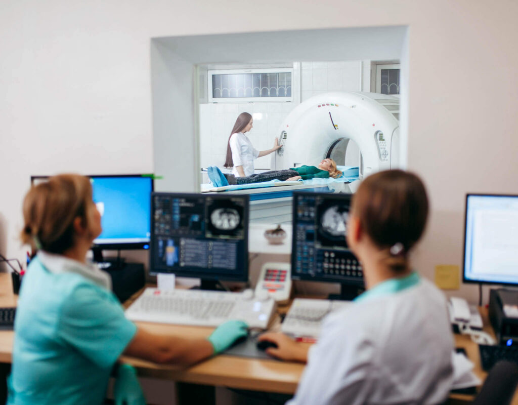
A DTI scan is performed as part of a brain MRI and usually takes 30–60 minutes.

What happens during your scan:
• Check-in and MRI safety screening
• Positioning with your head in a padded head coil
• Standard MRI sequences first
• DTI sequence lasting about 4–5 minutes
• Contrast injection if ordered

What you’ll experience:
• No pain
• Loud tapping or buzzing sounds
• Need to stay very still
• Ability to communicate with the technologist

After the scan, you can leave immediately with no recovery time.

What happens after my DTI scan is complete?

After your DTI scan is complete, you can leave immediately and resume normal activities.

Immediately after:
• Drive yourself home (unless sedated)
• Resume eating, drinking, and exercising
• No activity restrictions
• Drink water if contrast was used

Getting your results:
• Images reviewed by board-certified neuroradiologists
• Report sent to your referring doctor
• Scanslated® patient-friendly report sent via text within 48 hours

Your doctor will contact you to discuss findings and next steps.

When will I get my DTI scan results back?

Your doctor typically receives results within 24 hours. In urgent cases, preliminary results may be available sooner.

You will receive:
• A text message with a link to your imaging report
• Easy-to-understand explanations
• Anatomical diagrams

How accurate is a DTI scan?

DTI is highly accurate for detecting white matter abnormalities, brain connectivity problems, and neural pathway damage invisible on standard MRI.

Why DTI is highly accurate:
• Measures microscopic water diffusion
• Sensitive to subtle white matter changes
• Provides quantitative, objective data
• Detects damage before symptoms appear
• Tracks disease progression over time

DTI provides unique information unavailable from other imaging techniques when interpreted by experts.

How long does a DTI scan take?

A DTI scan typically takes 30–60 minutes total.

Time breakdown:
• Check-in and preparation: 10–15 minutes
• Standard brain MRI: 20–50 minutes
• DTI sequence: 4–5 minutes

How much does a DTI scan cost?

The cost of a DTI scan varies based on insurance coverage, whether contrast is used, and exam complexity.

American Health Imaging offers:
• In-network coverage with 99% of insurance plans
• Up to 60% cost savings compared to hospitals
• Transparent cost estimates before your visit
• Self-pay options for uninsured patients

Will my insurance cover a DTI scan?

Most insurance plans cover DTI scans when medically necessary and ordered by your physician, though coverage varies by plan.

Common covered indications:
• Brain tumor surgical planning
• Traumatic brain injury with persistent symptoms
• Stroke assessment and rehabilitation planning
• Multiple sclerosis diagnosis and monitoring
• Unexplained neurological symptoms
• Presurgical epilepsy evaluation
• Neurodegenerative disease assessment

American Health Imaging handles:
• Insurance verification
• Prior authorization
• Claims submission
• Cost transparency

Patient Reviews and Testimonials

With a 97% satisfaction score, we’re proud to deliver compassionate care, clear communication, and imaging you can count on.

Have you visited a American Health Imaging Center lately?

Your feedback helps us continue providing exceptional care. If you’ve had a recent experience at one of our locations, we’d love to hear from you.

Diffusion Tensor Imaging Articles and Resources

MRI for Seizures

Epilepsy and other types of seizure disorders are caused by structural changes in your brain. Magnetic resonance imaging, or MRI, offered at American Health Imaging

Read Article

Do you need to schedule a Diffusion Tensor Imaging?

American Health Imaging offers same-day and next-day appointment options to make getting an imaging scan convenient. Get a referral from your doctor, find the American Health Imaging center closest to you, and then call to schedule an imaging appointment.