NeuroQuant® Brain Imaging

What is NeuroQuant®?

NeuroQuant® is an advanced, FDA-cleared imaging analysis tool that enhances standard brain MRI by providing precise volumetric measurements of brain structures. This quantitative analysis helps physicians detect subtle changes in brain volume that may be associated with conditions such as dementia, Alzheimer’s disease, multiple sclerosis (MS), traumatic brain injury (TBI), and other neurologic disorders.

NeuroQuant® compares your brain structure volumes to a normative database based on age, sex, and cranial volume to identify abnormalities that could be difficult to detect on visual inspection alone. The detailed measurements support earlier diagnosis, more accurate treatment planning, and effective monitoring of neurological conditions over time.

NeuroQuant® is particularly helpful for:

  • Alzheimer’s disease and dementia evaluation
  • Multiple sclerosis (MS) monitoring
  • Traumatic brain injury (TBI) assessment
  • Cognitive impairment and memory concerns
  • Tracking brain changes over time with objective data
  • Earlier detection of neurodegenerative changes

NeuroQuant® Services at American Health Imaging

American Health Imaging is in-network with 99% of insurance plans and offers NeuroQuant® imaging with significant cost savings compared to hospital-based imaging.

NeuroQuant® Imaging Centers Near You

NeuroQuant® brain imaging services are available at most American Health Imaging locations throughout the Southeast and Texas. We offer same-day and next-day appointments with extended hours to match your schedule and needs.

Find Your Nearest NeuroQuant® Imaging Center

What is a NeuroQuant Scan Used For?

NeuroQuant® provides objective, age- and sex-adjusted measurements of brain anatomy that are valuable when evaluating patients with progressive neurological symptoms or cognitive changes. Volume measurements of regions such as the hippocampus, ventricles, and cortical structures help physicians identify brain atrophy, asymmetries, or other structural changes over time. American Health Imaging

Why Your Doctor May Recommend a NeuroQuant® Scan

  • Alzheimer’s disease and dementia
  • Multiple sclerosis (MS)
  • Traumatic brain injury (TBI)
  • Epilepsy and seizure disorders
  • Cognitive impairment or neurodegenerative symptoms
  • Monitoring brain changes over time

Types of NeuroQuant® Scans We Offer

A 1.2T High Field Open MRI is a type of Open MRI scanner that has a strong magnet (1.2 Tesla) and an open design, making it more comfortable for people who feel claustrophobic or have a larger body. The Open MRI has an open front and sides designed to provide increased comfort. The “high field” means it has a stronger magnetic field than some other MRI machines, which helps in getting more accurate images.

A 1.5T High Field MRI is a medical imaging machine that uses a strong magnetic field (1.5 Tesla) to create clear and detailed pictures of the inside of your body. The “high field” means it has a stronger magnetic field than some other MRI machines, which helps in getting more accurate images.

A 1.5T Wide Bore MRI is a type of medical imaging machine that uses a strong magnetic field (1.5 Tesla) to take detailed pictures of the inside of your body. The “wide bore” part means the opening of the machine is larger at 27 ½” wide, making it more comfortable for people who feel anxious or are larger in size. Many of our 1.5T Wide Bore MRIs feature faster scanning technology to reduce most exams to 15 minutes.

A 16-slice CT scanner is a medical imaging machine that takes detailed X-ray pictures of your body in thin slices, which are then combined to create a complete image. The “16-slice” part means it can capture 16 slices of images at once, allowing for faster and more detailed scans.

3D mammography, or tomosynthesis, is an advanced x-ray technology that takes multiple images of breast tissue to create a 3-dimensional picture of the breast. It differs from traditional mammography in that traditional mammography yields only a single image. A 3D image of the breast allows for better assessment of masses and reduces the likelihood of a false positive or an unnecessary biopsy.

A 3T MRI is a medical imaging machine that uses an even stronger magnetic field (3 Tesla) to take very detailed pictures of the inside of your body. Because of its high strength, it can capture clearer images and is often used for more complex scans. Often used for prostates and different types of brain imaging. The “wide bore” part means the opening of the machine is larger at 27 ½” wide, making it more comfortable for people who feel anxious or are larger in size.

A 64-slice CT scanner is a medical imaging machine that takes very detailed X-ray pictures of your body by capturing 64 slices of images at once. This allows for faster scans and even more detailed images, which is useful for diagnosing complex conditions. Our 64-slice CT features innovative technology that automates dose according to your size, weight, and anatomy, providing high-quality images with minimal radiation.

ABUS Breast Cancer Screening uses sound waves to create 3D pictures of the breast tissue. ABUS breast cancer screening technology is specifically developed and FDA-approved for women with dense breast tissue.

An arthrogram visualizes the inside of a joint, such as the shoulder, knee, hip, or wrist. A contrast dye is injected into the joint to make the joint structures, including ligaments, tendons, cartilage, and the joint capsule, more visible on X-ray or MRI images.

Coronary Calcium CT – also called Cardiac Scoring – measures calcified (hard) plaque inside the arteries that can grow and restrict blood flow to the muscles of the heart identifying potential Coronary Artery Disease (CAD) before you have symptoms.

A DEXA scan is a medical imaging technique used to measure bone mineral density. It is commonly performed to diagnose osteoporosis or assess the risk of developing fractures as we age.

Diffusion Tensor Imaging (DTI) is a special type of MRI technique that helps doctors see the pathways of nerve fibers in the brain. By looking at these images, doctors can better understand how the brain’s wiring works and diagnose conditions like brain injuries, tumors, or diseases that affect these connections.

Faster scanning MRI technology reduces the time patients spend on the table for scans by up to 50% to an average scan time of less than 15 minutes providing a more comfortable imaging experience for anxious patients or anyone in pain. AI technology delivers high quality images with reduced motion artifacts and noise distortions for the diagnostic insights providers need to determine next steps in patient care.

Fluoroscopy uses injected contrast dye and an X-Ray machine to take a continuous series of X-rays instead of individual snapshots. It is most commonly used to evaluate parts of your body that are moving in order to create a short video of your body system in motion. It is particularly useful for observing the digestive, urinary, respiratory, and reproductive systems and their functioning.

A screening mammography is a specialized X-ray of the breasts that takes images from different angles. These images can reveal abnormalities in breast tissues. This exam is annually recommended for all women over the age of 40.

Our MRI machines use a powerful magnetic field, radio waves, and a computer to generate detailed images of organs, tissues, bones, and other structures inside the body. With a larger bore or tunnel-like opening compared to traditional MRI machines, our wide-bore MRI provides high quality images without compromising patient comfort.

Our MRI machines use a powerful magnetic field, radio waves, and a computer to generate detailed images of organs, tissues, bones, and other structures inside the body. With a larger bore or tunnel-like opening compared to traditional MRI machines, our wide-bore MRI provides high quality images without compromising patient comfort.

Myelogram imaging is used to evaluate the spinal cord, nerve roots, and surrounding structures in the spine with an injectable contrast dye to enhance the visibility of the spinal cord and nerves on X-ray images.

Specialty post processing technology used for brain MRI to assess brain volume and atrophy for neurological diseases including dementia, Alzheimers, seizures, MS lesions and Traumatic Brain Injuries.

The Open Upright MRI, also known as a stand-up MRI, is the only MRI scanner able to scan you in multiple positions, including sitting, standing, bending (for flexion and extension) or lying down. This unique MRI provides natural weight-bearing imaging and is helpful for your doctor to diagnose the area where you experience pain. The Open Upright MRI is open in front of you, behind you, and above you. This open design may be more comfortable for people who feel anxious or are larger in size.

A PET/CT (Positron emission tomography) is a non-invasive exam that uses small amounts of radioactive materials called radiotracers to reveal how internal organs are functioning. The scan takes about 60 minutes and images are captured via CT as your body processes the radioactive material. This offers great insight into the root of the disease process or the source of symptoms.

What to Expect During Your NeuroQuant® Appointment

Your NeuroQuant® appointment starts with a standard brain MRI. Once the scan is complete, the NeuroQuant® software automatically analyzes your images to measure the volume of key brain structures, no extra scanning, no added steps, and nothing invasive. The MRI itself typically takes 30–60 minutes. You’ll lie comfortably on a cushioned table that slides into the scanner while images are captured, hearing rhythmic thumping and buzzing sounds that are completely normal. Earplugs or headphones are provided to keep you comfortable throughout the exam.

What makes NeuroQuant® convenient:

  • No additional radiation exposure—uses standard MRI technology
  • No special preparation beyond standard MRI requirements
  • Analysis performed automatically on your brain MRI images
  • Provides objective, quantitative data your doctor can track over time
  • Results typically available within 24-72 hours

Why Choose American Health Imaging for Your NeuroQuant®

American Health Imaging (AHI) is in-network with 99% of insurance plans for NeuroQuant® brain imaging and offers fast scheduling combined with advanced technology for accurate, comfortable exams.

  • Cost savings compared to hospital imaging
  • Advanced MRI scanners and optimized sequences for brain imaging
  • Board-certified radiologists with neuroimaging expertise
  • Flexible scheduling, including same-day and next-day appointments
  • Comfortable, outpatient settings with friendly staff

We offer convenient outpatient locations, efficient scheduling, and a supportive environment designed to reduce stress and wait times.

Schedule Your NeuroQuant® Exam

Scheduling your NeuroQuant® brain MRI with American Health Imaging is made easy in just a few steps:

  • Get a referral from your doctor (required for insurance coverage)
  • Choose your location from our imaging centers offering NeuroQuant® services
  • Schedule your appointment online or by phone—same-day slots often available
  • Verify insurance coverage—we’ll handle pre-approval and authorization
  • Follow standard MRI preparation instructions provided before your exam

Common Questions About NeuroQuant® Scans

What is NeuroQuant®?

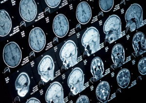
NeuroQuant® is an FDA-cleared software that provides precise volumetric measurements of brain structures by analyzing standard brain MRI images. It helps physicians detect and track subtle brain changes associated with neurological conditions that may not be visible through visual inspection alone.

How NeuroQuant® works:
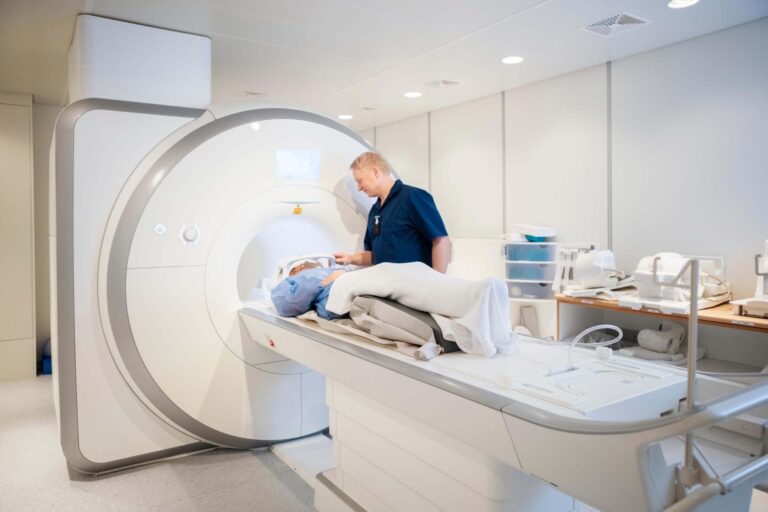
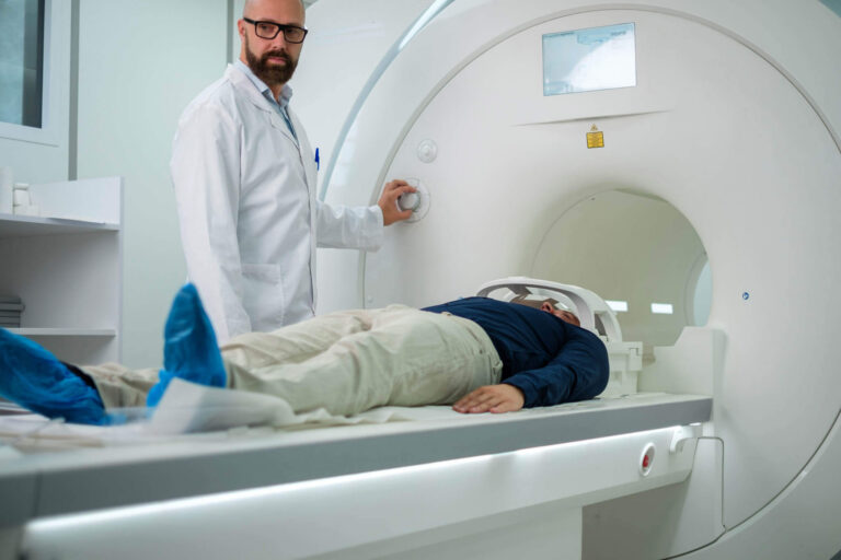
• You undergo a standard brain MRI scan
• NeuroQuant® software automatically analyzes your MRI images
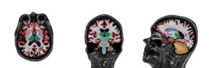
• Measures volumes of specific brain structures such as the hippocampus, ventricles, and cortical regions
• Compares your measurements to age-, sex-, and size-matched normative data
• Generates detailed reports highlighting areas of concern
• Provides objective data for tracking changes over time

NeuroQuant® is ideal for:
• Alzheimer’s disease and dementia evaluation
• Multiple sclerosis (MS) monitoring and treatment response
• Traumatic brain injury (TBI) assessment
• Cognitive impairment investigation
• Epilepsy and seizure disorder evaluation
• Establishing baseline measurements for future comparison

Key benefit: NeuroQuant® provides objective, quantitative measurements that help detect subtle brain changes earlier than visual assessment alone, supporting more timely diagnosis and treatment decisions.

Why did my doctor order a NeuroQuant® scan?

Your doctor ordered a NeuroQuant® scan because you need precise, objective measurements of brain structure volumes to help diagnose or monitor a neurological condition.

Common reasons for ordering NeuroQuant®:
• Memory loss or cognitive decline, including evaluation for Alzheimer’s disease or dementia
• Multiple sclerosis monitoring to assess brain atrophy and disease progression
• Traumatic brain injury to evaluate structural damage and recovery
• Establishing a baseline for comparison with future scans
• Monitoring whether treatments are slowing disease progression
• Diagnostic clarification when symptoms are unclear

The benefit: NeuroQuant® detects subtle volumetric changes in brain structures that may not be obvious on standard MRI images. For example, hippocampal atrophy is a key marker of Alzheimer’s disease that NeuroQuant® can precisely measure and track over time.

Do I need a referral for a NeuroQuant® scan?

Yes, you need a referral (also called a doctor’s order or prescription) to get a NeuroQuant® brain MRI.

What your referral does:
• Provides clinical information about your neurological symptoms or condition
• Is required for insurance coverage, including Medicare
• Ensures the exam is medically appropriate for your situation
• Allows for proper interpretation and follow-up

Once your doctor sends the order to American Health Imaging, we’ll contact you to schedule your appointment and verify your insurance coverage.

Schedule your NeuroQuant® exam:
https://americanhealthimaging.com/request-appointment/

How do I schedule a same-day NeuroQuant® scan?

Scheduling a same-day NeuroQuant® scan at American Health Imaging is easy and convenient.

Flexible scheduling options:
• Call us or request an appointment online
• Same-day and next-day appointments available at most locations
• Extended hours for maximum convenience
• Quick insurance verification process

Once your doctor sends us your referral, we can often schedule your brain MRI with NeuroQuant® analysis quickly at one of our convenient locations throughout the Southeast and Texas.

Schedule your NeuroQuant® exam:
https://americanhealthimaging.com/request-appointment/

What imaging centers near me offer NeuroQuant®?

American Health Imaging offers NeuroQuant® brain imaging at most locations across five states.

NeuroQuant® locations include:
• Alabama
• Georgia
• Tallahassee, FL
• San Antonio, TX
• South Carolina

NeuroQuant® is available at most American Health Imaging centers. To find the location nearest you or confirm services, use our online appointment request feature or call your preferred imaging center.

View all imaging centers:
https://americanhealthimaging.com/location/

What does a NeuroQuant® scan diagnose?

NeuroQuant® helps diagnose and monitor neurological conditions by measuring brain structure volumes and detecting abnormalities or changes over time.

Conditions evaluated with NeuroQuant®:
• Alzheimer’s disease (measures hippocampal volume loss)
• Dementia (evaluates patterns of brain atrophy)
• Mild cognitive impairment (MCI)
• Multiple sclerosis (tracks brain atrophy and treatment response)
• Traumatic brain injury (assesses structural damage)
• Epilepsy (evaluates hippocampal sclerosis)
• Normal pressure hydrocephalus (measures ventricular enlargement)

What NeuroQuant® measures:
• Hippocampus (critical for memory)
• Ventricles (fluid-filled spaces)
• Cortical gray matter
• White matter
• Specific brain lobes (frontal, temporal, parietal, occipital)

NeuroQuant® provides age-, sex-, and intracranial volume-adjusted percentiles to identify deviations from normal ranges.

How should I prepare for my NeuroQuant® scan?

Preparing for a NeuroQuant® scan is the same as preparing for a standard brain MRI and requires minimal preparation.

Before your appointment:
• No fasting required—eat and drink normally
• Continue taking regular medications unless advised otherwise
• Inform us of any metal implants, devices, or foreign bodies
• Tell us if you have claustrophobia or anxiety
• Arrange transportation if you will receive sedation

Day of your appointment:
• Wear comfortable, loose-fitting clothing without metal fasteners
• Remove all jewelry, watches, hairpins, and metal accessories
• Bring your insurance card and photo ID
• Bring a list of current medications

What should I expect during a NeuroQuant® scan?

During your NeuroQuant® exam, you’ll undergo a standard brain MRI while lying comfortably in the scanner. The NeuroQuant® analysis happens automatically after your scan—no additional imaging is required.

What happens during your scan:
• Check-in and MRI safety screening
• Removal of metal objects and changing into a gown if needed
• Positioning on a cushioned table with your head in a special coil
• Earplugs or headphones provided for comfort
• MRI scanning for 30–60 minutes
• Communication with the technologist via intercom

The NeuroQuant® analysis occurs after you leave and does not add time to your appointment.

What happens after my NeuroQuant® scan is complete?

After your NeuroQuant® brain MRI, you can resume all normal activities immediately.

Immediately after your scan:
• Resume driving and daily activities
• No dietary restrictions
• No recovery time or side effects

Getting your results:
• NeuroQuant® software analyzes your MRI images
• Board-certified neuroradiologists review both the MRI and volumetric data
• Your doctor receives a comprehensive report within a few hours
• You receive a patient-friendly report via text within 48 hours

Your doctor will discuss findings and next steps with you.

When will I get my NeuroQuant® results?

Your doctor typically receives results within a few hours after the report is completed.

You will receive:
• A text message with a link to your imaging report within 48 hours

The report includes clear explanations, diagrams, and information to help prepare for your follow-up appointment.

How accurate is NeuroQuant®?

NeuroQuant® is highly accurate and reliable for measuring brain structure volumes and is FDA-cleared with extensive clinical validation.

Accuracy highlights:
• Precise, reproducible volumetric measurements
• Removes subjectivity from visual MRI interpretation
• Validated against manual segmentation (the gold standard)
• Uses large normative databases for comparison
• Excellent for tracking changes over time

NeuroQuant® is particularly accurate for hippocampal volume measurement, a key marker in Alzheimer’s disease.

How long does a NeuroQuant® scan take?

The MRI scanning portion takes 30–60 minutes.

Total appointment time:
• 45–75 minutes including check-in and setup

The NeuroQuant® analysis is performed automatically after your scan and does not require additional time in the scanner.

How much does a NeuroQuant® scan cost?

The cost of a NeuroQuant® brain MRI depends on insurance coverage and individual benefits.

Typical costs:
• With insurance: Standard copay, coinsurance, or deductible
• Significant savings compared to hospital-based imaging
• NeuroQuant® analysis is often included with the MRI

American Health Imaging verifies coverage, obtains authorization, and provides a transparent cost estimate before your appointment.

Will my insurance cover a NeuroQuant® scan?

Most insurance plans, including Medicare, cover medically necessary brain MRI exams. NeuroQuant® analysis is typically included when ordered for appropriate clinical indications.

Generally covered for:
• Dementia or cognitive decline evaluation
• Alzheimer’s disease assessment
• Multiple sclerosis monitoring
• Traumatic brain injury evaluation
• Neurological symptoms requiring brain imaging

American Health Imaging handles insurance verification and prior authorization.

Is NeuroQuant® safe?

Yes. NeuroQuant® is extremely safe.
Safety facts:
• No radiation exposure
• Non-invasive—no needles or procedures
• No additional scanning beyond standard MRI
• No contrast dye typically required
• FDA-cleared technology

NeuroQuant® itself has no risks. Standard MRI safety screening applies, and our team reviews all safety considerations before your exam.

Patient Reviews and Testimonials

With a 97% satisfaction score, we’re proud to deliver compassionate care, clear communication, and imaging you can count on.

Have you visited a American Health Imaging Center lately?

Your feedback helps us continue providing exceptional care. If you’ve had a recent experience at one of our locations, we’d love to hear from you.

NeuroQuant® Articles and Resources

Do you need to schedule a NeuroQuant®?

American Health Imaging offers same-day and next-day appointment options to make getting an imaging scan convenient. Get a referral from your doctor, find the American Health Imaging center closest to you, and then call to schedule an imaging appointment.