Ultrasound Imaging (Sonography)

What is an Ultrasound?

Ultrasound, also called sonography, is a safe, painless, and non-invasive imaging technique that uses high-frequency sound waves to create real-time images of the inside of your body. Unlike X-rays and CT scans, ultrasound uses no radiation, making it one of the safest imaging modalities available. The technology works on the same principle as sonar used by submarines and echolocation used by bats.

During an ultrasound exam, a handheld device called a transducer is placed on your skin and emits sound waves that bounce off internal structures, such as organs, blood vessels, and soft tissues. These echoes are captured and instantly transformed by a computer into detailed moving images displayed on a screen.

Ultrasound is commonly used to diagnose and monitor:

  • Abdominal organ conditions (liver, gallbladder, kidneys, pancreas, spleen)
  • Vascular problems and blood flow
  • Musculoskeletal injuries (tendons, ligaments, muscles, joints)
  • Soft tissue masses and cysts
  • Thyroid and other glandular conditions
  • Guidance for biopsies and medical procedures

Ultrasound Services at American Health Imaging

American Health Imaging is in-network with 99% of insurance plans and offers ultrasound services up to 60% less than hospital-based imaging.

Ultrasound Imaging Centers Near You

Find convenient ultrasound imaging near you at any of our American Health Imaging centers. We offer same-day and next-day appointments with extended hours and weekend availability to match your schedule and needs.

Find Your Nearest Ultrasound Imaging Center

What is an Ultrasound Used For?

Ultrasound imaging helps doctors visualize soft tissues and fluid-filled structures inside your body to diagnose diseases, monitor conditions, and guide medical procedures, all without radiation exposure. Ultrasound is particularly effective at distinguishing solid masses from fluid-filled cysts, a task that’s surprisingly difficult with other imaging techniques.

Common Ultrasound Exams and Uses:

  • Abdominal Ultrasound: Checks the liver, gallbladder, pancreas, spleen, kidneys, and major vessels; helps detect stones, liver disease, cysts, tumors, and abdominal pain causes.
  • Renal (Kidney) Ultrasound: Evaluates kidney size and structure; identifies stones, blockages, infections, and monitors chronic kidney disease or transplants.
  • Vascular Ultrasound (Doppler): Measures blood flow in arteries and veins; detects clots, narrowed vessels, aneurysms, and circulation issues.
  • Musculoskeletal Ultrasound: Images muscles, tendons, ligaments, joints, and nerves; used for sports injuries, tears, inflammation, and guiding injections.
  • Thyroid Ultrasound: Assesses thyroid nodules, enlargement, inflammation, and helps guide biopsies.
  • Pelvic Ultrasound: Examines the bladder, uterus, and ovaries; evaluates pelvic pain, bleeding, cysts, fibroids, and masses.
  • Breast Ultrasound: Helps investigate lumps or mammogram findings and distinguishes cysts from solid masses.

Types of Ultrasound Scans We Offer

A 1.2T High Field Open MRI is a type of Open MRI scanner that has a strong magnet (1.2 Tesla) and an open design, making it more comfortable for people who feel claustrophobic or have a larger body. The Open MRI has an open front and sides designed to provide increased comfort. The “high field” means it has a stronger magnetic field than some other MRI machines, which helps in getting more accurate images.

A 1.5T High Field MRI is a medical imaging machine that uses a strong magnetic field (1.5 Tesla) to create clear and detailed pictures of the inside of your body. The “high field” means it has a stronger magnetic field than some other MRI machines, which helps in getting more accurate images.

A 1.5T Wide Bore MRI is a type of medical imaging machine that uses a strong magnetic field (1.5 Tesla) to take detailed pictures of the inside of your body. The “wide bore” part means the opening of the machine is larger at 27 ½” wide, making it more comfortable for people who feel anxious or are larger in size. Many of our 1.5T Wide Bore MRIs feature faster scanning technology to reduce most exams to 15 minutes.

A 16-slice CT scanner is a medical imaging machine that takes detailed X-ray pictures of your body in thin slices, which are then combined to create a complete image. The “16-slice” part means it can capture 16 slices of images at once, allowing for faster and more detailed scans.

3D mammography, or tomosynthesis, is an advanced x-ray technology that takes multiple images of breast tissue to create a 3-dimensional picture of the breast. It differs from traditional mammography in that traditional mammography yields only a single image. A 3D image of the breast allows for better assessment of masses and reduces the likelihood of a false positive or an unnecessary biopsy.

A 3T MRI is a medical imaging machine that uses an even stronger magnetic field (3 Tesla) to take very detailed pictures of the inside of your body. Because of its high strength, it can capture clearer images and is often used for more complex scans. Often used for prostates and different types of brain imaging. The “wide bore” part means the opening of the machine is larger at 27 ½” wide, making it more comfortable for people who feel anxious or are larger in size.

A 64-slice CT scanner is a medical imaging machine that takes very detailed X-ray pictures of your body by capturing 64 slices of images at once. This allows for faster scans and even more detailed images, which is useful for diagnosing complex conditions. Our 64-slice CT features innovative technology that automates dose according to your size, weight, and anatomy, providing high-quality images with minimal radiation.

ABUS Breast Cancer Screening uses sound waves to create 3D pictures of the breast tissue. ABUS breast cancer screening technology is specifically developed and FDA-approved for women with dense breast tissue.

An arthrogram visualizes the inside of a joint, such as the shoulder, knee, hip, or wrist. A contrast dye is injected into the joint to make the joint structures, including ligaments, tendons, cartilage, and the joint capsule, more visible on X-ray or MRI images.

Coronary Calcium CT – also called Cardiac Scoring – measures calcified (hard) plaque inside the arteries that can grow and restrict blood flow to the muscles of the heart identifying potential Coronary Artery Disease (CAD) before you have symptoms.

A DEXA scan is a medical imaging technique used to measure bone mineral density. It is commonly performed to diagnose osteoporosis or assess the risk of developing fractures as we age.

Diffusion Tensor Imaging (DTI) is a special type of MRI technique that helps doctors see the pathways of nerve fibers in the brain. By looking at these images, doctors can better understand how the brain’s wiring works and diagnose conditions like brain injuries, tumors, or diseases that affect these connections.

Faster scanning MRI technology reduces the time patients spend on the table for scans by up to 50% to an average scan time of less than 15 minutes providing a more comfortable imaging experience for anxious patients or anyone in pain. AI technology delivers high quality images with reduced motion artifacts and noise distortions for the diagnostic insights providers need to determine next steps in patient care.

Fluoroscopy uses injected contrast dye and an X-Ray machine to take a continuous series of X-rays instead of individual snapshots. It is most commonly used to evaluate parts of your body that are moving in order to create a short video of your body system in motion. It is particularly useful for observing the digestive, urinary, respiratory, and reproductive systems and their functioning.

A screening mammography is a specialized X-ray of the breasts that takes images from different angles. These images can reveal abnormalities in breast tissues. This exam is annually recommended for all women over the age of 40.

Our MRI machines use a powerful magnetic field, radio waves, and a computer to generate detailed images of organs, tissues, bones, and other structures inside the body. With a larger bore or tunnel-like opening compared to traditional MRI machines, our wide-bore MRI provides high quality images without compromising patient comfort.

Our MRI machines use a powerful magnetic field, radio waves, and a computer to generate detailed images of organs, tissues, bones, and other structures inside the body. With a larger bore or tunnel-like opening compared to traditional MRI machines, our wide-bore MRI provides high quality images without compromising patient comfort.

Myelogram imaging is used to evaluate the spinal cord, nerve roots, and surrounding structures in the spine with an injectable contrast dye to enhance the visibility of the spinal cord and nerves on X-ray images.

Specialty post processing technology used for brain MRI to assess brain volume and atrophy for neurological diseases including dementia, Alzheimers, seizures, MS lesions and Traumatic Brain Injuries.

The Open Upright MRI, also known as a stand-up MRI, is the only MRI scanner able to scan you in multiple positions, including sitting, standing, bending (for flexion and extension) or lying down. This unique MRI provides natural weight-bearing imaging and is helpful for your doctor to diagnose the area where you experience pain. The Open Upright MRI is open in front of you, behind you, and above you. This open design may be more comfortable for people who feel anxious or are larger in size.

A PET/CT (Positron emission tomography) is a non-invasive exam that uses small amounts of radioactive materials called radiotracers to reveal how internal organs are functioning. The scan takes about 60 minutes and images are captured via CT as your body processes the radioactive material. This offers great insight into the root of the disease process or the source of symptoms.

What to Expect During Your Ultrasound Appointment

Your ultrasound is a simple, comfortable step in taking charge of your health. You’ll start by getting settled on the exam table (or seated, depending on the area being scanned). A warm, water‑based gel is applied to your skin to help create clear images. The technologist then glides a small handheld device over the area, using gentle pressure to capture detailed views. You may be asked to shift positions or briefly hold your breath so we can get the best angles.

Ultrasounds are painless for most patients, though you may feel mild pressure if you’re being scanned for an injury or have a full bladder. Images appear instantly and are saved for the radiologist to review, keeping you informed and empowered at every step.

Typical ultrasound appointment:

  • Check-in and paperwork: 10-15 minutes
  • Exam duration: 15-45 minutes (depending on complexity)
  • Total appointment time: 30-60 minutes

Every step is designed to keep you comfortable, informed, and confident—because staying proactive about your health makes you a true wellness warrior.

Why Choose American Health Imaging for Your Ultrasound

American Health Imaging (AHI) is in-network with 99% of insurance plans for ultrasound imaging and offers fast scheduling combined with experienced technologists for accurate, comfortable exams.

  • Significant cost savings up to 60% compared to hospital ultrasound pricing
  • Experienced, ARDMS-certified sonographers who specialize in ultrasound imaging
  • Same-day and next-day appointments available with convenient scheduling
  • Extended and weekend hours at many outpatient imaging centers
  • Board-certified radiologists with subspecialty training provide accurate interpretations
  • Real-time imaging allows dynamic evaluation during your exam
  • No radiation exposure—completely safe for repeat imaging
  • With a 97% patient satisfaction score, patients love our compassionate techs and comfortable environment

Schedule Your Ultrasound Today

Getting started is easy:

  • Get a referral from your doctor (required for insurance coverage)
  • Choose your location from our centers across Alabama, Georgia, Florida, Texas, and South Carolina
  • Schedule your appointment online or by phone—same-day slots often available
  • Follow preparation instructions—we’ll provide specific guidance if your exam requires fasting or a full bladder

Common Questions About Ultrasound Scans

How do I schedule my ultrasound?

Once your doctor determines that an ultrasound is necessary, their office will typically send us your prescription or order, and we’ll contact you to schedule your appointment. We verify your insurance coverage and obtain any required pre-authorizations.

If you don’t have insurance or have a high deductible, we also offer affordable self-pay rates.

We offer same-day and next-day appointments to ensure your exam fits your schedule.

Schedule your ultrasound:
https://americanhealthimaging.com/request-appointment/

View all imaging centers:
https://americanhealthimaging.com/location/

Do I need a referral for an ultrasound?

Yes, you need a referral (also called a doctor’s order) to get an ultrasound.

What your referral does:
• Specifies which body area needs to be scanned
• Indicates whether contrast is needed
• Is required for insurance coverage
• Ensures the right exam is performed for your specific medical concerns

Once your doctor sends the order to American Health Imaging, we’ll contact you to schedule your appointment and verify your insurance coverage.

Schedule your ultrasound:
https://americanhealthimaging.com/request-appointment/

How should I prepare for my ultrasound appointment?

Preparation for ultrasound varies depending on the type of exam you’re having. Your doctor or our scheduling team will provide specific instructions when your appointment is scheduled.

Typical preparation may include:

Abdominal ultrasound (liver, gallbladder, pancreas, spleen):
• Eat a fat-free meal the evening before
• Fast (no food or drink except water) for 8–12 hours
• Prevents gas buildup and improves image clarity

Renal (kidney) ultrasound:
• Drink 4–6 glasses (32–48 ounces) of water 1 hour before
• Do NOT empty your bladder
• A full bladder improves visualization

Pelvic ultrasound:
• Drink 32 ounces of water 1 hour before
• Arrive with a full bladder
• Do NOT empty your bladder

Musculoskeletal ultrasound:
• No special preparation required
• Wear loose-fitting clothing

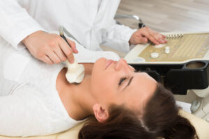
Thyroid/neck ultrasound:
• No special preparation required
• Avoid necklaces or turtlenecks

General tips:
• Arrive 10–15 minutes early
• Bring photo ID and insurance card
• Bring a list of medications
• Inform the technologist of symptoms or concerns

What should I expect during my ultrasound appointment?

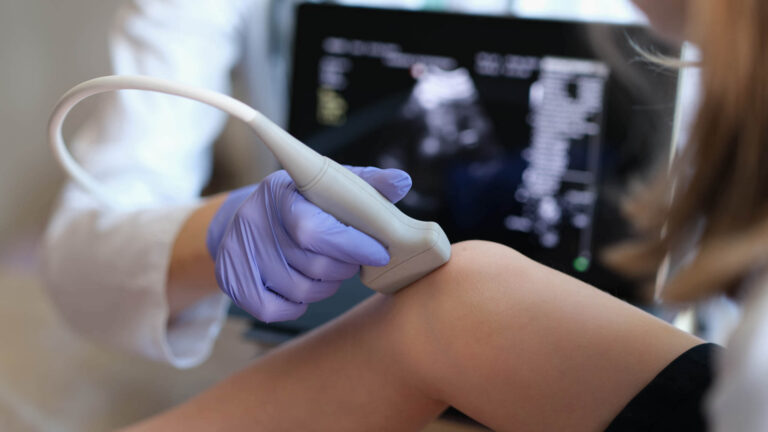
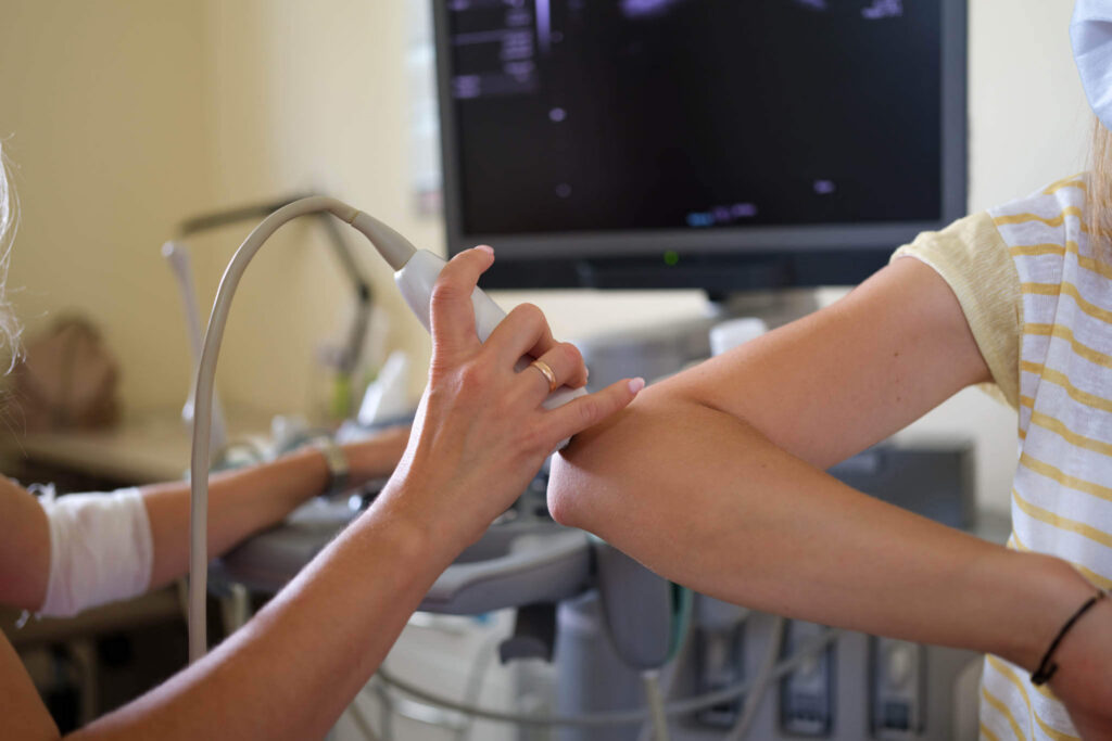
Your ultrasound exam is simple, comfortable, and usually painless.

What happens during your exam:
• You may change into a gown depending on the area being examined
• You’ll lie on an exam table (sometimes seated or standing)
• Warm gel is applied to your skin
• The technologist moves a handheld transducer over the area
• You may be asked to change positions or briefly hold your breath
• Doppler exams may produce whooshing sounds
• Most exams take 15–45 minutes

You may feel mild pressure from the transducer. If you’re uncomfortable or have a full bladder, let the technologist know.

Does ultrasound use radiation?

No. Ultrasound uses absolutely no radiation. It relies only on high-frequency sound waves, similar to sonar.

Ultrasound is ideal for patients who:
• Need repeated imaging
• Want to avoid radiation exposure
• Require frequent monitoring
• Cannot undergo CT due to contrast allergies or kidney issues

Because there is no radiation, ultrasound can be safely performed as often as medically necessary.

How long does an ultrasound take?

The length of your ultrasound depends on the type of exam.

Typical exam times:
• Focused ultrasound: 15–20 minutes
• Abdominal ultrasound: 30–45 minutes
• Musculoskeletal ultrasound: 20–30 minutes
• Vascular Doppler study: 30–60 minutes
• Thyroid ultrasound: 15–20 minutes
• Renal ultrasound: 20–30 minutes

Total appointment time, including check-in, is usually 30–60 minutes.

What should I expect after my ultrasound appointment?

You can resume all normal activities immediately after your ultrasound.

After your exam:
• No recovery time
• No side effects
• Eat, drink, drive, and exercise normally
• Use the restroom immediately if you had a full bladder

Getting your results:
• Board-certified radiologists review your images
• Your doctor typically receives results within a few hours
• You receive a patient-friendly report via text within 48 hours

Why am I asked to have a full bladder for my ultrasound exam?

A full bladder is important for pelvic and kidney ultrasounds.

Why it helps:
• Improves sound wave transmission
• Pushes bowel gas out of the way
• Acts as a landmark for pelvic organs
• Provides contrast for clearer images

Not all ultrasounds require a full bladder. We’ll let you know when scheduling if this preparation is necessary.

Why can’t I eat or drink before my abdominal ultrasound?

Fasting improves image quality for abdominal ultrasounds.

Why fasting matters:
• Prevents gas buildup that blocks sound waves
• Keeps the gallbladder full and visible
• Reduces organ movement
• Prevents imaging artifacts

You may still:
• Take essential medications with small sips of water

Only certain abdominal exams require fasting. Kidney, pelvic, musculoskeletal, and thyroid ultrasounds usually do not.

Can I get my ultrasound results from the technologist?

No. Ultrasound technologists are not permitted to interpret results or provide diagnoses.

What the technologist can do:
• Perform the exam
• Capture high-quality images
• Explain the procedure
• Answer process-related questions

What they cannot do:
• Interpret findings
• Diagnose conditions
• Discuss abnormalities

Only a board-certified radiologist can interpret your images and send results to your doctor.

How much will my ultrasound cost?

Ultrasound costs vary depending on the exam type and body area.

Factors affecting cost:
• Type of ultrasound
• Complexity of the exam
• Doppler blood flow evaluation
• Insurance deductible status
• Hospital vs. outpatient imaging

American Health Imaging offers transparent pricing up to 60% less than hospital ultrasound costs.

Will my insurance cover my ultrasound?

Yes, most insurance plans cover medically necessary ultrasounds ordered by your doctor.

American Health Imaging:
• In-network with 99% of insurance plans
• Verifies coverage and obtains pre-authorization
• Provides cost estimates before your exam

If you don’t have insurance, affordable self-pay options are available.

What are the advantages of ultrasound over CT or MRI?

Ultrasound offers several unique advantages:

Advantages:
• No radiation exposure
• Real-time imaging
• Dynamic evaluation during movement
• Excellent at distinguishing fluid from solid masses
• Less expensive than MRI or CT
• No contrast dye required
• No claustrophobia
• Safe with metal implants and pacemakers
• Can guide procedures in real time

Other imaging may be better when:
• MRI is needed for detailed soft tissue or brain imaging
• CT is needed for bones, lungs, or bleeding

Is ultrasound safe? Are there any risks?

Yes. Ultrasound is extremely safe with no known risks or side effects.

Why ultrasound is safe:
• Uses sound waves only
• Non-invasive
• No cumulative exposure risk
• Safe for all ages

The only discomfort may be temporary pressure from the transducer or a full bladder.

What can an ultrasound show? What can’t it show?

What ultrasound shows well:
• Soft tissue organs
• Fluid-filled structures
• Blood flow with Doppler
• Muscles, tendons, ligaments, and joints
• Gallstones and kidney stones
• Real-time movement

What ultrasound doesn’t show well:
• Bones
• Air-filled organs like lungs
• Deep structures behind bone
• Brain tissue

Your doctor may recommend additional imaging if needed.

Can ultrasound detect cancer?

Ultrasound can detect masses that may represent cancer, but it cannot definitively diagnose cancer without a biopsy.

Ultrasound can:
• Identify solid vs. cystic masses
• Detect suspicious characteristics
• Guide biopsies
• Monitor tumor size

Biopsy and additional imaging are often required for confirmation.

How quickly will I get my ultrasound results?

Your doctor typically receives results within a few hours after the report is finalized.

You will receive:
• A text message with a link to your patient-friendly report within 48 hours

What is musculoskeletal ultrasound?

Musculoskeletal (MSK) ultrasound images muscles, tendons, ligaments, joints, and nerves.

MSK ultrasound can diagnose:
• Tendon tears
• Ligament injuries
• Muscle strains
• Bursitis
• Nerve compression
• Joint inflammation

Advantages:
• Real-time dynamic imaging
• No metal interference
• Immediate assessment
• Lower cost than MRI

Can ultrasound detect gallstones and kidney stones?

Yes. Ultrasound is the best test for detecting gallstones and is often the first imaging test ordered.

Gallstones:
• Sensitivity of 95% or higher
• Detects inflammation and bile duct issues

Kidney stones:
• Detects most stones
• Excellent for identifying blockages
• CT is better for very small stones

Ultrasound is preferred for pregnant patients, children, and frequent monitoring.

Do I need to remove my clothes for an ultrasound?

It depends on the exam and your clothing.

You may keep clothes on if:
• The area is easily accessible
• Clothing doesn’t interfere

You’ll change into a gown if:
• Having abdominal, pelvic, or kidney ultrasound
• Clothing blocks the area

Wearing loose, two-piece clothing is recommended.

Can I eat after my ultrasound if I had to fast before?

Yes. You can eat and drink normally immediately after your ultrasound.

You can also:
• Take medications
• Drive yourself home
• Return to work or exercise

There is no recovery time.

Why did my doctor order an ultrasound instead of an MRI or CT?

Your doctor ordered ultrasound because it’s often the best first imaging test.

Ultrasound is ideal when:
• Evaluating fluid vs. solid masses
• Assessing blood flow
• Avoiding radiation
• Quick results are needed
• Cost is a consideration
• Metal implants are present

MRI or CT may be ordered if more detailed imaging is required.

Patient Reviews and Testimonials

With a 97% satisfaction score, we’re proud to deliver compassionate care, clear communication, and imaging you can count on.

Have you visited a American Health Imaging Center lately?

Your feedback helps us continue providing exceptional care. If you’ve had a recent experience at one of our locations, we’d love to hear from you.

Ultrasound Articles and Resources

Do you need to schedule a Ultrasound?

American Health Imaging offers same-day and next-day appointment options to make getting an imaging scan convenient. Get a referral from your doctor, find the American Health Imaging center closest to you, and then call to schedule an imaging appointment.