
If you are experiencing pain in the shoulder, you know how limiting it can be to our daily lives. If you have a painful shoulder injury, and your healthcare provider thinks an MRI scan is appropriate, it may be because they think you could have a torn rotator cuff.
A doctor will often order a shoulder MRI to diagnose rotator cuff injuries because it offers the best available imaging scan for finding out whether you have a torn rotator cuff. Let’s explore why MRI scans are ideal for this purpose and what they reveal about your shoulder.
What causes a torn rotator cuff?
A torn rotator cuff occurs when one or more tendons detach from the bone, either partially or completely. This could result from trauma, gradual wear and tear, or repetitive overhead motion. Something as simple as painting, swimming, or tennis could contribute to this injury.
How does an MRI create images of the shoulder?
When you have a shoulder injury, it helps your provider to know exactly what kind of injury has happened, and where.
An MRI uses a strong magnetic field and radio waves to reveal detailed images of soft tissues and bones within the shoulder. This advanced imaging technology shows your provider the type, exact location, and severity of a shoulder injury.
What can an MRI scan reveal about my shoulder?
An MRI scan provides the most detailed imaging available to reveal the health of your shoulder and surrounding tissues.
An MRI scan offers unparalleled detail, highlighting:
- Wear and tear of the tendon, including partial and full-thickness injuries.
- The condition of muscle tissue, pinpointing shrinkage or weakening caused by chronic injury.
- Cartilage, which can point to any infection or potential arthritis in the joint causing your pain..
- Ligament injuries, bone abnormalities, and nerve compression.
- Nerve damage and nerve compression.
These insights ensure your provider can develop an accurate diagnosis and treatment plan.
How does the level of detail in MRI imaging help diagnose a shoulder injury?
MRI scans help to diagnose shoulder injuries by providing detailed and comprehensive information about the soft tissues, bones, and joint structures. An MRI will reveal location, size, and severity of tears in rotator cuff tendons, as well as nearby tissues.
The imaging precision seen on an MRI scan allows providers to:
- Identify tears, strains, or inflammation in tendons, muscles, and ligaments.
- Detect even minor tears or early tissue degeneration not visible on other scans.
How can an MRI show the condition of the tendons, muscles, and ligaments in my shoulder?
An MRI provides a detailed look at the tendons, muscles, and ligaments in your shoulder, allowing your healthcare provider to assess not only the structure but also the overall health of these soft tissues. Your MRI results will help your provider to identify any areas that show overstress, weakness, or damage.
An MRI scan can detect subtle injuries (like small tears, inflammation, tendon fraying, or fluid buildup) that might otherwise go unnoticed. It also helps distinguish between partial and complete tears, showing their exact size and location. Additionally, an MRI can reveal signs of chronic wear or damage, providing valuable insights into both recent and long-term injuries.
Why MRI scans are ideal for diagnosing a torn rotator cuff
MRI scans are the preferred method to diagnose shoulder injuries, including a torn rotator cuff, because they are non-invasive and do not use radiation. An MRI will also show an assessment of the surrounding structures and conditions of your shoulder joint.
A torn rotator cuff might mean a small tear in a tendon connecting to the bone (a partial tear), or it could mean a full-thickness rupture (a complete tear), which is a complete separation of the tendon from the bone. An MRI provides the level of detail to distinguish between the type and severity of such an injury.
Why is an MRI a great choice for identifying rotator cuff injuries?
An MRI provides the best available imaging for a detailed look at tendons in the shoulder joint. The scan detects even small tears within the rotator cuff, showing the severity and location of any injuries or abnormalities.
Surrounding structures and tissues, like ligaments, muscles, and cartilage, are also evaluated to determine if any other injuries or weaknesses might be present. On MRI results, each of these soft tissues looks a little different from the other tissues.
How does an MRI distinguish between a partial tear and a complete tear in the rotator cuff?
MRI scans distinguish between a partial tear, where the tendon remains partially attached, and a complete tear, where it is fully separated.
- Partial tears appear as fraying or thinning of the tendon on an MRI, but it is still partially attached.
- Complete tears show a clear separation, often with fluid buildup or muscle atrophy. This often creates a visible gap or detraction.
The distinction between these two presentations provides additional clues about the tear’s severity and how long it’s been present.
Inflammation, swelling, and degeneration: what MRI scans reveal
An MRI scan can detect inflammation, swelling, and degeneration in your shoulder by capturing detailed images of the soft tissues and surrounding structures.
- Inflammation often appears as increased fluid or swelling, which shows up clearly on an MRI.
- Degeneration, on the other hand, might look like thinning, fraying, or irregular texture in the tendons and other tissues, indicating chronic wear or aging.
- MRI results can also highlight subtle changes in tissue composition, like scar tissue formation or reduced blood supply, which are common in long-term injuries.
How MRI scans help guide treatment for a torn rotator cuff
With the detailed images from an MRI scan, your provider can:
- Diagnose the size, location, and severity of the tear.
- Recommend conservative treatments like physical therapy or anti-inflammatory medications for partial tears.
- Plan surgical repair for severe tears or cases involving tendon retraction.
By offering a comprehensive view of your injury, the MRI ensures personalized and effective care.
What details about a torn rotator cuff will my MRI results offer my healthcare provider?
Your MRI results will show your healthcare provider detailed information about your torn rotator cuff, including whether the tear is partial-thickness (incomplete separation of the tendon from the bone) or more severe full-thickness (complete separation of the tendon from the bone).
Additional details may include the size, nature, and location of the tear, along with any inflammation or swelling that is present, including in the tissue surrounding your shoulder.
How does an MRI help differentiate rotator cuff tears from other potential causes of shoulder pain?
An MRI helps differentiate rotator cuff tears from other causes of shoulder pain by providing detailed images of not only the tendons but also the surrounding muscles, ligaments, cartilage, and joint structures.
Shoulder pain can stem from various issues, like bursitis, tendonitis, labral tears, or even arthritis, all of which present differently on an MRI. An MRI can also identify fluid buildup, swelling, or structural abnormalities that might not be linked to a tendon tear.
If I have a torn rotator cuff, how will my provider use my MRI results to create a treatment plan?
Your healthcare provider will use your MRI results to understand the size, location, and severity of your rotator cuff tear, as well as any associated issues like inflammation or tendon degeneration.
If the tear is small or partial, they might recommend conservative treatments such as physical therapy, rest, or anti-inflammatory medications to restore strength and mobility. For larger or complete tears, especially if the tendon has retracted or if there’s significant muscle damage, surgical repair may be necessary.
The MRI also helps your provider anticipate potential complications, like poor tendon healing or long-term weakness, and adjust your treatment plan accordingly. By offering a clear picture of your injury, the MRI ensures your care is personalized and targeted for the best possible outcome.
What to expect during your shoulder MRI
There are a few things to keep in mind for the day of your MRI scan to help diagnose a torn rotator cuff. We’ll walk you through the whole process, including how to prepare, what happens during your scan, and what to expect from your MRI results.
How should I prepare for a shoulder MRI?
Be sure to follow your healthcare provider’s instructions in preparing for the scan. They’ll create a tailor-made set of instructions for your specific condition, and for your specific scan, to ensure that you get accurate results.
To get ready for your shoulder MRI, you will want to remove all metal, like jewelry, watches, and piercings, since the metal will interfere with the magnetic field. You will also want to inform your provider if you have any metal implants in your body that may affect the scan, and if you have allergies to iodine or other contrast agents if contrast dye will be used.
What should I expect during my upcoming shoulder MRI scan?
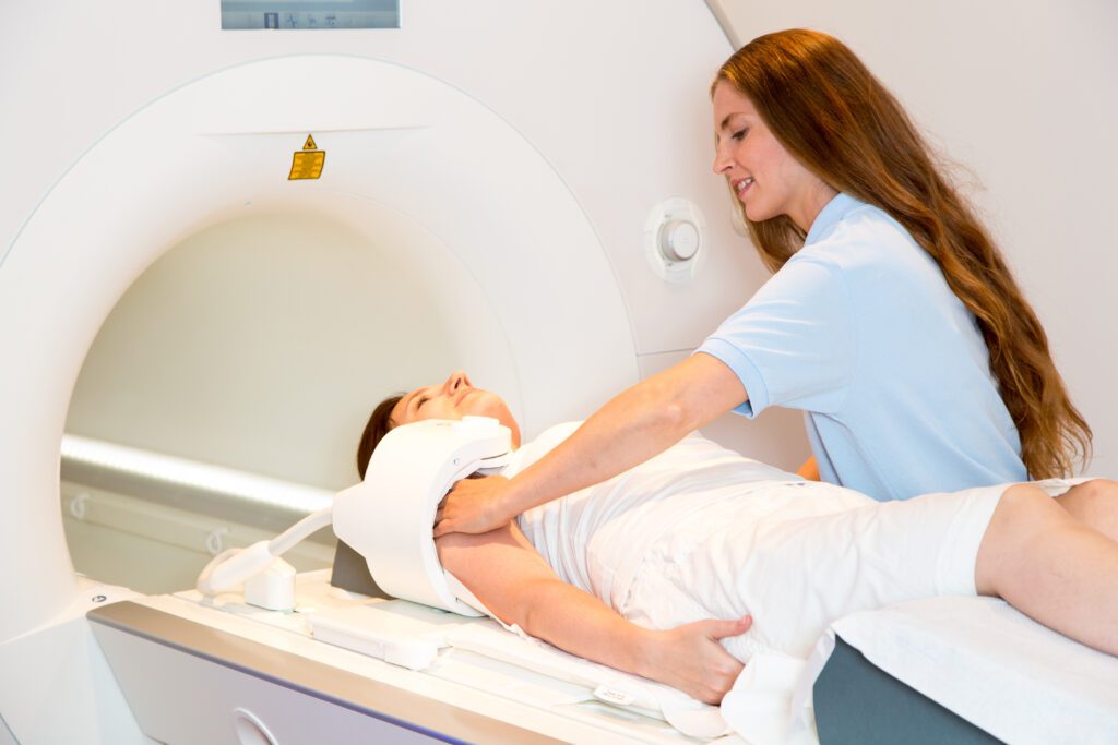
For your shoulder MRI scan, you may be asked to change into a gown. You will lie down on a bed-like table that will move in and out of the MRI machine to capture detailed images of your shoulder.
You will be asked to lie still during the scan, to keep the resolution of images as sharp as possible. You may be asked to hold your breath for brief intervals of a couple of seconds throughout the scan.
During the scan, you may be given earplugs or headphones, as the machine makes clanging or tapping noises as it takes images. Your MRI scan will take between 30 minutes to an hour, depending on your circumstances.
How do we ensure your comfort during an MRI?
At American Health Imaging, we prioritize your comfort during every step of your MRI scan. Our technologists are highly trained to position your shoulder in a way that minimizes discomfort while ensuring the best quality imaging results.
You’ll have constant communication with your technologist via an intercom system inside the MRI machine, and they can see and hear you throughout the scan. If you’re feeling nervous or anxious, let them know—they’re there to support you and make your experience as stress-free as possible.
What is fast scan technology?
We understand that imaging can feel overwhelming, especially when you’re experiencing pain. That’s why we are proud to offer advanced scanning technology that reduces scan times by up to 50%, making the process quicker and more comfortable.
With our fast scan technology, your shoulder MRI could take less than 10 minutes, helping you get the answers you need without disrupting your day.
How to schedule a shoulder MRI appointment with us
Reach out to us at American Health Imaging, and we’ll help you schedule an appointment at an imaging center near you, today.
We’re here to help you get the answers you need.
Frequently Asked Questions About Shoulder MRIs
Q: How does an MRI create images of the shoulder?
A: MRI uses powerful magnets and radio waves to create detailed cross-sectional images of the shoulder’s structures.
Q: What can an MRI scan reveal about my shoulder?
A: An MRI can show detailed images of muscles, tendons, ligaments, cartilage, and bone, highlighting injuries or abnormalities.
Q: How does the level of detail in MRI imaging help diagnose a shoulder injury?
A: The high-resolution images allow healthcare providers to precisely locate and assess injuries, ensuring accurate diagnosis and treatment.
Q: Why is MRI an excellent choice for identifying rotator cuff injuries?
A: MRI provides unparalleled detail of soft tissues, making it ideal for detecting damage to the rotator cuff’s tendons and muscles.
Q: How does an MRI distinguish between a partial and a complete rotator cuff tear?
A: MRI imaging can show the severity of tendon damage, helping your provider differentiate between a minor tear and a full rupture.
Q: What can an MRI show about inflammation or degeneration in my shoulder?
A: MRI scans can reveal swelling, fluid buildup, or tissue degeneration that might contribute to shoulder pain or dysfunction.
Q: How does an MRI help differentiate rotator cuff tears from other shoulder problems?
A: The scan’s detail allows providers to rule out conditions like arthritis, bursitis, or other injuries that may mimic a rotator cuff tear.
Q: How should I prepare for a shoulder MRI?
A: You’ll wear comfortable clothing for your scan, remove any metal objects you’re wearing, and inform the technologist of any implants you may have.