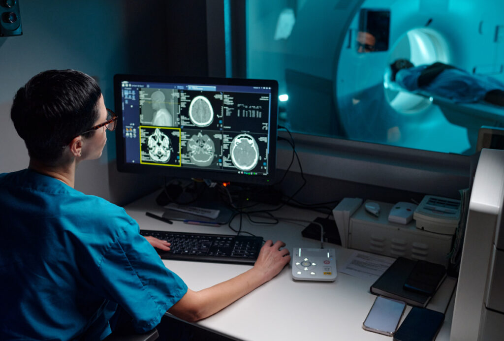
If you’ve been dealing with tingling, numbness or a burning pain that keeps coming back, your doctor may recommend an MRI to find out what is causing it. An MRI can show many nerve problems, especially those caused by compression, inflammation, injury or growths pressing on a nerve. It captures detailed images of your nerves and the tissue around them to help your doctor find the source of your symptoms.
In this guide, you’ll learn which nerve problems appear on an MRI, how accurate the scan is, what symptoms are worth checking and how your results guide the next step in your care.
What Is an MRI?
An MRI, short for “magnetic resonance imaging,” is a safe and non-invasive way to see what’s happening inside your body. The scanner combines powerful magnets, radio waves and computer processing to build detailed pictures of organs, soft tissue, bone and nerves. The scanner captures those pictures in thin slices, so your radiologist can look at one small section of your body at a time.
MRI is especially useful for nerve pain because it shows small, soft structures like nerves in greater detail than many other imaging tests. An MRI also uses no radiation, which can be helpful if your doctor needs to image the same area more than once. Before your scan, your imaging team will complete a quick safety screening for implants or metal.
What Nerve Problems Does an MRI Show?
Nerve pain can have several different causes, so you may need an MRI to help find out what is causing your symptoms. Here are the main nerve problems an MRI can show and what they may look like on the images.
Pinched or Compressed Nerves
An MRI can detect a pinched nerve, which happens when nearby tissue presses on a nerve and interrupts the signals traveling through it. The images can show exactly what is creating that pressure. Possible causes include a herniated or bulging disc, a bone spur, a narrowed spinal canal, thickened ligaments or swelling in the surrounding tissue.
The nerve itself can also show signs of pressure on an MRI. A compressed nerve root may look flattened, pushed out of position or slightly swollen just past the point of pressure.
Because an MRI shows each level of your spine in detail, your doctor can see which nerve is under pressure and where that pressure starts. For example, pain that travels down your leg may come from a compressed nerve in your lower back.
Nerve Inflammation
When a nerve becomes inflamed, it holds more fluid than healthy tissue. That extra fluid appears as a bright area on the MRI images, so an irritated nerve stands out even when nothing is pressing on it. An inflamed nerve may also look thicker than the matching nerve on the other side of your body.
An MRI scan helps your doctor identify conditions such as optic neuritis, which affects the nerve behind the eye, or inflammation in the brachial plexus, the bundle of nerves that runs from your neck through your shoulder and into your arm. Contrast dye makes active inflammation even easier to spot, because inflamed areas take up the dye and light up on the scan.
Nerve Damage
Nerve damage looks different from nerve irritation. After an injury, a nerve may appear thinned, interrupted or surrounded by scar tissue. An MRI can show whether an injury stretched a nerve, partly tore it or cut through it completely. Your doctor needs that detail before deciding whether the nerve can heal on its own or needs surgical repair.
An MRI also picks up changes in the muscles a nerve controls. A muscle that stops receiving normal signals may show fluid-like changes at first, then shrink over time. Those changes can point to nerve damage and give your doctor an idea of how long it’s been happening.
Tumors or Masses Affecting Nerves
Sometimes a growth is the reason a nerve isn’t working the way it should. An MRI is the leading tool for finding tumors, cysts and other masses along a nerve. Some grow from the nerve sheath, which is the protective covering around a nerve, while others form nearby and press inward.
Your radiologist can measure a mass, see which structures it touches and see how far it extends. Contrast dye often makes the edges of a growth even easier to define. Finding a mass early means you and your doctor can decide on your next treatment sooner.
What Neurological Conditions Don’t Show on an MRI?
An MRI shows the physical structure of your nerves and everything around them, including their shape, size and condition. A few nerve conditions affect how a nerve sends signals rather than how it looks, so doctors confirm those with nerve function testing.
Conditions that may not appear on an MRI include:
- Small fiber neuropathy, which affects the tiniest nerve endings in the skin
- Early diabetic peripheral neuropathy
- Nerve symptoms linked to chemotherapy
- Neuropathy caused by a vitamin deficiency
- Widespread pain conditions such as fibromyalgia
- Some cases of carpal tunnel syndrome, where the nerve looks normal, but signals travel slowly
Getting an MRI to rule out certain conditions is still an important step. It shows your doctor whether a disc, tumor or narrowed space is behind your symptoms. Based on those results, your doctor may also recommend a nerve conduction study, which measures how well signals travel along your nerves, or an electromyogram (EMG), which measures electrical activity in your muscles.
What Are the Symptoms of Nerve Damage?
The symptoms you experience depend on which type of nerve is affected, and you may notice several kinds of symptoms at the same time:
- Sensory nerves carry touch, temperature and pain signals. Damage can cause numbness, tingling, burning or electric shock sensations, trouble sensing hot and cold or difficulty keeping your balance.
- Motor nerves control the muscles you move. Damage can lead to weakness, cramping, twitching, muscle loss or trouble gripping objects and buttoning a shirt.
- Autonomic nerves manage automatic functions such as digestion, heart rate and sweating. Damage can cause blood pressure changes, lightheadedness, sweating too much or too little, nausea, bloating or bladder changes.
Symptoms can build slowly over years or come on within hours. Nerves heal slowly, so finding the cause of your nerve problems early gives you the best chance at a full recovery. If your symptoms aren’t improving, talk with your doctor about whether imaging can help. Sudden weakness, numbness on one side of your body, or new loss of bladder or bowel control needs emergency care right away.
Is a CT Scan or MRI Better for Nerve Damage?
Doctors use MRI and CT scans for different reasons. A CT scan takes multiple images of the body and is especially useful for showing bone detail, fractures and bleeding. Some CT exams also produce 3D views that give your doctor even greater insight.
For nerve damage, an MRI is uniquely positioned to help. It separates nerves from the muscle, fat and fluid around them and picks up the small signal changes that point to inflammation or injury.
For example, a CT scan may show a bone spur crowding a nerve. An MRI shows the spur and how the nerve is responding to it. If you can’t have an MRI because of an implant or device, your doctor may recommend a myelogram to get a detailed look at the spinal canal and nerve roots.
Can an MRI See Nerve Damage Without Contrast?
An MRI can show nerve damage without contrast. It can reveal compressed nerve roots, herniated discs, narrowing in the spinal canal and muscle changes linked to long-term nerve damage.
Your doctor may recommend contrast when they need a closer look at inflammation, a possible tumor or scar tissue from a previous surgery. If your MRI requires contrast, a technologist will give it to you through a small IV during the exam.
Schedule an MRI With American Health Imaging
If your doctor recommends an MRI to find the cause of your nerve pain, American Health Imaging can help you get clear answers. We use advanced MRI technology and explain each step of the exam as you go. Most of our locations offer wide-bore MRI, and open MRI is available at select centers if you feel anxious in tight spaces. A board-certified, subspecialized radiologist reads your exam, and you’ll receive a Scanslated® report by text within 48 hours after your report is finalized. Your report uses plain language and diagrams, so you can understand your results before your follow-up appointment with your doctor.
American Health Imaging is in-network with 99% of major insurance plans, offers imaging at up to 60% less than hospital imaging and has a 97% patient satisfaction rating.
Talk to your doctor about imaging and schedule your MRI with American Health Imaging today.
Frequently Asked Questions About Getting MRIs for Nerve Problems
MRI is the preferred scan for showing nerve damage. It provides the clearest view of nerves and the soft tissue around them, and it can reveal compression, inflammation, injury and masses in a single exam.
An MRI can show structural nerve problems with a high degree of accuracy. Very small nerve fibers may be too tiny to see on an MRI or other imaging test. Even so, your MRI can help your doctor determine whether pressure, inflammation or a growth may be causing your symptoms.
Most nerve MRIs don’t require contrast. Your doctor may order it to look for active inflammation, get a clearer view of a tumor or distinguish scar tissue from healthy nerve tissue after surgery.
A normal MRI can still give your doctor useful information by showing that there’s no obvious structural problem causing your symptoms, such as a herniated disc, spinal narrowing or mass. Your doctor may then recommend nerve function testing or blood work to look for other possible causes.
Most MRI exams take 30 to 60 minutes, depending on the area being scanned and how many images your doctor needs. American Health Imaging's faster scanning MRI technology, available at many of our locations, reduces many scan times to as little as 8 minutes.
You will find American Health Imaging centers in five states, with MRI available in Alabama, Georgia, Tallahassee, FL, Beaumont, TX, San Antonio, TX and South Carolina. Once your provider sends a referral, you can book online or by phone at the center closest to you. Same-day appointments are available, and extended and weekend hours are offered at select locations.