

Digital X-ray (also called digital radiography or DR) is a modern imaging technique that uses digital sensors instead of traditional photographic film to capture X-ray images of the inside of your body. Like the difference between film cameras and digital cameras, digital X-rays produce instant, high-quality images that can be viewed immediately on a computer screen.
Key benefits:
• Less radiation than traditional film X-rays
• Clearer, more detailed images
• Quick and painless—typically 5–10 minutes total
• Instant results—no waiting for film development
• Environmentally friendly—no chemicals needed
Digital X-rays are commonly used to diagnose:
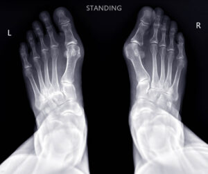
• Bone fractures and breaks
• Joint dislocations and arthritis
• Chest conditions (pneumonia, lung infections, heart problems)
• Dental problems
• Digestive system issues
• Infections and inflammation
• Foreign objects in the body
Your doctor ordered a digital X-ray to see inside your body and diagnose injuries, detect diseases, or monitor existing conditions. X-rays are excellent for visualizing bones and detecting many common medical problems.
Common reasons your doctor ordered this test:
• Bone and joint concerns
• Chest and lung evaluation
• Abdominal and digestive issues
Other reasons:
• Unexplained persistent pain without an obvious cause
• Pre-surgical evaluation
• Monitoring chronic conditions
• Routine screening (such as chest X-rays for certain occupations)
The benefit:
X-rays provide immediate, clear images that help your doctor make accurate diagnoses and create effective treatment plans quickly and affordably.
Scheduling a same-day digital X-ray at American Health Imaging is easy once you have your doctor’s referral.
Flexible scheduling options:
• Call your preferred American Health Imaging location directly
• Request an appointment online through our website
• Same-day and next-day appointments available at most locations
• Extended evening and weekend hours at select centers
Important:
Digital X-ray services are available only at select American Health Imaging locations in Alabama, Georgia, and Texas. When you call to schedule, confirm that your preferred center offers digital X-ray exams.
View imaging centers:
https://americanhealthimaging.com/location/
Yes, you need a referral (also called a doctor’s order or prescription) to get a digital X-ray at American Health Imaging.
What your referral does:
• Specifies which body part needs to be X-rayed
• Provides relevant clinical information about your symptoms
• Is required for insurance coverage
• Ensures the correct exam is performed for your medical needs
Once your doctor sends the order to American Health Imaging, we’ll contact you to schedule your appointment and verify your insurance coverage.
Schedule your X-ray:
https://americanhealthimaging.com/request-appointment/
American Health Imaging offers digital X-ray services at convenient locations near you in Alabama, Georgia, and Texas, including locations in San Antonio and Beaumont.
To find the imaging center nearest you:
• Call your desired imaging center to confirm availability
• Use our online appointment request
• Ask about convenient appointment times
All digital X-ray centers offer up to 60% cost savings compared to hospital-based imaging and are in-network with 99% of insurance plans.
View all imaging centers:
https://americanhealthimaging.com/location/
Digital X-rays require minimal preparation.
Before your appointment:
• Wear comfortable, loose-fitting clothing without metal zippers, buttons, or snaps
• Remove jewelry before leaving home if possible
• Bring your insurance card and photo ID
• Bring any relevant previous X-rays
At your appointment:
• Arrive 10–15 minutes early for paperwork
• Remove all metal objects (jewelry, watches, belts, hairpins, hearing aids, eyeglasses, dentures with metal, bras with underwire)
• Change into a gown if your clothing has metal
• Remove cell phones and keys from pockets
Tell the technologist if you:
• Are pregnant or might be pregnant
• Have metal implants or joint replacements
• Cannot hold the required position due to pain
• Have had recent X-rays of the same area
Some specialized X-rays may require fasting or contrast—specific instructions will be provided if needed.
Your digital X-ray appointment is quick and painless.
What happens:
• Check-in and insurance verification
• Safety screening for pregnancy and metal implants
• Changing into a gown if needed
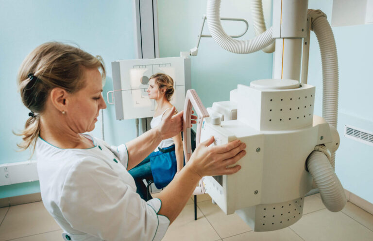
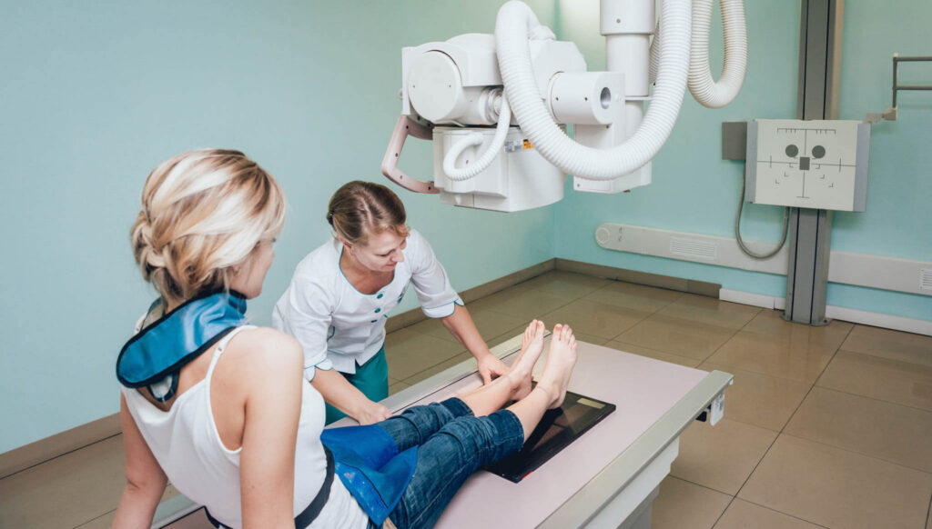
• Positioning (sitting, standing, or lying down)
• Lead shielding for areas not being imaged
• Remaining still for 2–5 seconds per image
• Brief breath-holding for chest X-rays
• Image review to ensure quality
Most exams include 2–4 images and take 5–10 minutes total.
The actual X-ray imaging takes just 5–10 seconds per image, with most exams requiring 2–4 images.
Typical appointment times:
• Single area (hand, foot, elbow): 10–15 minutes
• Chest X-ray: 10–15 minutes
• Spine or multiple areas: 15–25 minutes
You can leave immediately after your exam—no recovery time is needed.
You can resume all normal activities immediately after your digital X-ray.
After your exam:
• No side effects from the X-ray itself
• Temporary soreness may occur if an injured area was positioned
Getting your results:
• Images reviewed by board-certified radiologists
• Report sent to your doctor
• Patient-friendly report sent via text
Your doctor will contact you to discuss results and next steps.
Digital X-ray costs vary depending on the body area being imaged and how many views are needed. X-rays are generally one of the most affordable imaging tests.
Factors affecting cost:
• Body area being imaged
• Number of views required
• Insurance deductible status
• Hospital vs. outpatient imaging center
American Health Imaging offers transparent pricing up to 60% less than hospital X-ray charges.
Yes, most insurance plans cover medically necessary X-rays ordered by your doctor.
American Health Imaging is in-network with 99% of insurance plans, meaning lower out-of-pocket costs for you. We verify coverage, obtain prior authorization if needed, and provide a cost estimate before your exam.
If you don’t have insurance, affordable self-pay options are available.
Yes, digital X-rays are very safe. The radiation exposure from a single X-ray is extremely small and comparable to the natural background radiation you receive over a few days or weeks.
Digital X-rays use 50–80% less radiation than traditional film X-rays while producing higher-quality images. Modern equipment and trained technologists follow the ALARA principle (As Low As Reasonably Achievable) to minimize exposure.
Digital and traditional X-rays both use X-ray radiation, but they differ in how images are captured and processed.
Digital X-ray:
• Captured on digital sensors
• Instant results on a computer screen
• 50–80% less radiation
• Images can be enhanced and shared electronically
• No film or chemical processing
• Environmentally friendly
Traditional film X-ray:
• Captured on photographic film
• Requires chemical development
• Higher radiation exposure
• Fixed image quality
• Physical film transport and storage
Digital X-rays have largely replaced film X-rays due to better quality, safety, and convenience.
X-rays during pregnancy require careful consideration. Always tell your doctor and technologist if you are pregnant or might be pregnant.
Important facts:
• Most diagnostic X-rays expose the fetus to extremely low radiation
• No single diagnostic X-ray exceeds accepted safety limits
• Chest and extremity X-rays pose minimal risk
• Abdominal and pelvic X-rays are avoided unless medically necessary
Doctors will only perform X-rays during pregnancy when the medical benefit outweighs any theoretical risk.
Yes, digital X-rays are safe for children when medically necessary. Pediatric X-rays use lower radiation doses adjusted for the child’s size and age.
Pediatric safety measures:
• Adjusted radiation dosing
• Lead shielding
• Fast imaging to reduce motion
• Pediatric-trained technologists
• Parents may stay in the room with shielding
Common pediatric X-rays include chest X-rays, fracture evaluation, and swallowed object detection.
What digital X-rays show well:
• Bones and fractures
• Joint alignment and arthritis
• Dense objects (metal, swallowed items, kidney stones)
• Air-filled structures like lungs
• Heart size and shape
• Certain lung conditions
What digital X-rays do not show well:
• Soft tissues (muscles, tendons, ligaments)
• Brain tissue
• Spinal cord
• Blood vessels
• Early-stage soft tissue cancers
Your doctor may recommend MRI, CT, or ultrasound if more detail is needed.
Yes. You can request copies of your digital X-ray images and radiology report from the American Health Imaging location where your exam was performed.
How to access your report:
• Receive a text message with a Scanslated® link
• View patient-friendly explanations
• Review results before seeing your doctor
Copies are helpful for second opinions, specialist visits, or personal medical records.
Your doctor ordered an X-ray because it is often the best first imaging test.
X-rays are ideal when:
• Bone fractures or breaks are suspected
• Quick results are needed
• Bone and joint issues are the primary concern
• Cost is a consideration
• Lower radiation exposure is preferred
MRI or CT may be ordered when soft tissue detail or more complex imaging is required.
Both X-ray and fluoroscopy use X-ray technology, but they serve different purposes.
X-ray (radiography):
• Static images
• Brief exposure
• Lower radiation dose
• Used for bones, joints, chest
Fluoroscopy:
• Real-time moving images
• Continuous X-ray beam
• Higher radiation dose
• Used during medical procedures
Think of X-rays as photographs and fluoroscopy as an X-ray video.
If you move during the X-ray exposure, the image may be blurred and need to be repeated.
If this happens:
• The technologist sees it immediately
• The image is repeated at no extra charge
• Positioning adjustments are made
Tips to stay still:
• Communicate discomfort
• Follow breath-hold instructions
• Use supports if needed
Modern digital X-rays are very fast, making it easier to stay still.
For most digital X-rays, you can eat and drink normally.
No fasting required for:
• Bone X-rays
• Chest X-rays
• Joint X-rays
Some specialized exams may require fasting, such as abdominal X-rays or studies using contrast. We’ll provide instructions if your exam requires special preparation.