
Your healthcare provider may recommend a brain MRI scan if they want to take a closer look at the condition of your brain, to see how it compares to the brain of someone who has epilepsy. A brain MRI scan creates detailed images of your brain, capturing cross-sectional views (pictures), so your healthcare provider can clearly see every part of your brain.
For people being evaluated for epilepsy, a MRI scan is particularly useful for identifying brain changes that could be causing seizures. These MRI images can give your healthcare provider important information about any potential sources of seizure activity, and as necessary, MRI images can help guide further steps toward your treatment.
We’ll show you how a brain MRI can help diagnose epilepsy, how an MRI scan detects changes in your brain and the cause of seizures, and why your provider may recommend additional imaging. Let’s find out more.
What is a brain MRI scan? How does it work?
An MRI is a type of imaging scan that uses powerful magnets and radio waves to create detailed pictures of your brain. This allows your healthcare provider to see different parts of your brain with exceptional clarity, highlighting any brain changes that might be present, including brain changes that may be the cause of epileptic seizures.
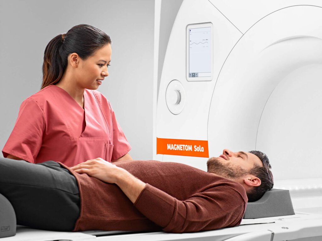
During a brain MRI, you’ll lie down on a table that slides into a large, doughnut-shaped machine. You might hear some loud noises as the machine takes the images, but don’t worry, this is completely normal. The whole scan takes about 30 minutes to an hour, depending on your provider’s orders, and on your circumstances.
Why do healthcare providers recommend a brain MRI for diagnosing epilepsy?
When it comes to diagnosing epilepsy, a brain MRI can help your healthcare provider identify any structural changes in the brain that could be contributing to seizures. By using your MRI results to pinpoint these changes, your healthcare provider can better understand the underlying cause of your seizures, which is important for developing an effective treatment plan.
In some cases, a brain MRI can also help your healthcare provider to determine the type of epilepsy you might have, so they can choose the most appropriate treatment plan for you. Also, your MRI results can help your provider rule out other potential causes of seizures, ensuring that your condition is diagnosed as accurately as possible.
Detecting the brain changes associated with epilepsy
Brain MRI scans are particularly useful at finding the source of any brain changes related to the onset of epilepsy. We’ll show you the brain changes an MRI can find, how an MRI finds changes associated with epileptic seizures, and how those brain changes are connected to epilepsy.
What types of brain changes are commonly associated with epilepsy?
Some of the most common brain changes associated with epilepsy include scarring from past brain injuries, abnormal blood vessels, and developmental malformations. These brain changes can sometimes interfere with the normal electrical activity in the brain, leading to seizures.
Other possible brain changes include tumors, infections that have caused inflammation, and changes in the brain’s structure due to stroke or other conditions. These issues might not always be visible to the naked eye, but they can often be detected using a brain MRI.
How does a brain MRI detect brain lesions or other changes?
A brain MRI uses strong magnets and radio waves to produce detailed images of your brain. These images can show even the smallest changes in brain tissue, allowing your healthcare provider to spot any brain changes or lesions that could be related to epilepsy.
Brain lesions, which are areas of damaged tissue, might not always cause noticeable symptoms, but they can disrupt the brain’s normal electrical activity and lead to seizures.
MRI images are incredibly detailed, and clearly show different types of tissues in the brain, allowing your provider to examine highly detailed images of your brain. This high level of detail helps your healthcare provider see the exact location, size, and shape of any abnormalities, which is crucial for understanding how a brain change might be contributing to seizures.
How can these brain changes help in determining the cause of epilepsy?
Identifying brain changes through an MRI can provide valuable clues about the cause of epilepsy. Similarly, finding a brain tumor or abnormal blood vessel can indicate that the seizures are a result of these specific structural changes. This information is essential for making a more accurate diagnosis and determining the most effective treatment approach.
Understanding the specific brain changes associated with epilepsy helps healthcare providers develop a treatment plan that targets the underlying cause of the seizures, rather than just managing the symptoms. This approach can lead to better outcomes and a higher quality of life for those living with epilepsy.
Using an MRI to find the source of seizure activity
To find the source of epileptic seizures, your healthcare provider will examine your brain MRI report results, which can even help predict future seizures. Let’s look at how a brain MRI locates the source of seizures, why finding the source of seizures is so important, and how your MRI results can indicate potential seizure activity in the future.
How does a brain MRI help my provider find the source of seizure activity?
The high-resolution images from an MRI can reveal brain changes that might be triggering seizures, such as scar tissue, tumors, or areas where brain cells may have developed differently. Your healthcare provider can use your MRI images to identify specific regions that appear abnormal and may be linked to your seizure activity. In some cases, the MRI can detect subtle changes in the brain that other imaging techniques might miss.
Why is it important to pinpoint the location of any seizure activity?
Knowing the exact source of seizures allows your healthcare provider to tailor treatment specifically to your needs. Also, understanding the precise location of seizure activity can help in predicting the type of seizures you might experience, and how your condition might evolve over time. This information is vital for developing a long-term management plan, whether it involves medication, lifestyle adjustments, or other therapeutic options.
How does an MRI scan show brain changes that could cause seizures in the future?
An MRI scan can show changes in the brain that might increase the risk of seizures in the future. These changes might include areas of the brain that are prone to scarring, regions with abnormal blood vessel formation, or parts of the brain that have developed differently due to genetic factors. By identifying these potential issues early, your healthcare provider can better anticipate future seizure activity, and adjust your treatment plan accordingly.
Follow-up diagnostic imaging for epilepsy
After analyzing your brain MRI results, and discussing them with you, your healthcare provider may recommend additional diagnostic imaging. We’ll show you why your results may indicate that you need further imaging, how follow-up imaging could support your care, and why your provider recommended a follow-up scan.
How does my provider use my MRI results to guide further testing or treatment decisions?
Your healthcare provider uses your brain MRI results to make informed decisions about what steps to take next in your diagnosis and treatment plan. If your provider identifies any areas of concern, such as scar tissue, abnormal growths, or structural changes, they may recommend additional imaging, just to get more information. Based on what the MRI reveals, your provider might suggest specific treatments tailored to your specific condition.
How can follow-up imaging help my healthcare provider to monitor the brain changes related to epilepsy?
Follow-up imaging is crucial in monitoring how your brain may change over time, especially if you have been diagnosed with epilepsy, or if you are at risk of developing it. Regular imaging allows your healthcare provider to track any progressions or new developments in your brain that could influence your condition. This ongoing monitoring helps your provider understand how effective your current treatments are, and whether any adjustments are needed.
After my MRI, why did my provider recommend additional diagnostic imaging for epilepsy?
Your provider may recommend additional diagnostic imaging after your MRI to get a more comprehensive view of your brain and its function. While an MRI provides detailed images of the brain’s structure, other types of imaging can offer additional insights into how your brain is working.
These scans can help identify areas of abnormal electrical activity or assess changes that might not be visible on an MRI. Additional imaging can also help clarify any findings from your MRI that may need further investigation. Ultimately, combining different types of imaging gives your healthcare provider a fuller picture of your brain health, so they can have the information they need to offer you the best care.
How to schedule a MRI appointment with us
Reach out to us at American Health Imaging, and we’ll help you schedule an appointment at an imaging center near you, today.
We’re here to help you get the answers you need.

Frequently Asked Questions
Q: What is a brain MRI scan and how does it work?
A: A brain MRI scan uses magnetic fields and radio waves to create detailed images of the brain, to help your provider to detect brain changes.
Q: Why is a brain MRI recommended for diagnosing epilepsy?
A: A brain MRI helps identify brain changes that might be causing seizures, aiding in accurate diagnosis and treatment planning.
Q: What types of brain changes are commonly associated with epilepsy?
A: Brain changes such as lesions, tumors, or structural changes are often linked to epilepsy, and show up on MRI results.
Q: How does a brain MRI detect brain lesions or changes?
A: An MRI produces high-resolution images that can reveal lesions, scars, or malformations in brain tissue.
Q: How can identifying brain changes help in determining the cause of epilepsy?
A: Finding brain changes can help pinpoint the specific cause of epilepsy, which is essential for choosing the right treatment.
Q: How does a brain MRI help my provider find the source of seizure activity?
A: A brain MRI can show the exact location of changes that may be causing seizures, helping to determine their origin.
Q: Why might a provider recommend additional diagnostic imaging after an MRI?
A: Additional imaging might be needed if the MRI reveals changes, or if more detail is required to plan treatment.
Q: How can follow-up imaging help monitor brain changes related to epilepsy?
A: Follow-up imaging allows healthcare providers to track any progression of brain changes, and adjust treatment as necessary.